Supported By


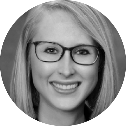
Kourtney Houser, MD
Dr. Houser is an Assistant Professor of Ophthalmology at the University of Tennessee in Memphis.
1. Please share with us your background.
I grew up in eastern Tennessee and attended the University of Tennessee in Knoxville for college. I studied biomedical engineering and originally wanted to pursue a career in materials science or biomechanics. I fell in love with more patient-centered care when volunteering at a hospital in town, and I decided to switch directions and attend medical school at the University of Tennessee Health Science Center. It was a scary decision at the time, as there are no physicians in my family, but I am so thankful that I went down this path. I was lucky enough to stay at the University of Tennessee for ophthalmology residency and then go on to Baylor for a fellowship in cornea, anterior segment, and refractive surgery.
2. What drew you to ophthalmology and, specifically, to your field of interest?
My engineering background initially drew me to ophthalmology—I loved the constant technological advances in the field as well as the complexity of ocular physiology. Once I shadowed in the field, I saw the immense influence that ophthalmologists have on patients’ lives by improving, maintaining, or enhancing vision and by diagnosing and monitoring systemic diseases. The intricacies of cornea and refractive surgery drew me into my subspecialty. We have so many new and exciting technologies and techniques that are constantly evolving and improving our care of patients.
3. Please describe your current position.
I am an Assistant Professor of Ophthalmology at Hamilton Eye Institute at the University of Tennessee Health Science Center, where I am thankful to have a practice with a wide range of pathology in cornea, anterior segment, and refractive surgery. I have the privilege of working with and learning from residents every day while helping them grow into knowledgeable and talented physicians and surgeons.
4. Who are your mentors?
The biggest blessing I have had in my career is the long list of wonderful mentors that have invested in me. All of my faculty and coresidents at the University of Tennessee supported me and taught me how to become a surgeon and physician. I also had the immense honor of training with Douglas Koch, MD; Marshall Hamill, MD; Stephen Pflugfelder, MD; Mitchell Weikert, MD; Sumitra Khandelwal, MD; and Zaina Al-Mohtaseb, MD, at Baylor for fellowship. They patiently molded me into the surgeon and person I am today and continue to support me and my career.
5. What has been the most memorable experience of your career thus far?
The most memorable moments in my career are times when I have been able to take the skills that I have been fortunate to obtain through the instruction of others and use them to treat and connect with patients. Most recently, my favorite experience was treating a cognitively delayed patient with keratoconus and a corneal perforation who was initially referred to me by another cornea specialist. With the help of my amazing staff, I was able to calm the patient enough to glue her perforation in clinic and then care for her through her corneal transplant. She is now always so excited to see me and my staff, and she gives us the biggest hugs when she comes in for follow-up.
I have also had the privilege of caring for multiple patients who have been supported by our local Lion’s Club. These patients have had significant cataracts, keratoconus, or other ocular pathology limiting their ability to work and care for themselves and no financial means to pay for intervention. Participating in their care and giving them their independence back has been a true blessing.
6. What are some new technological advances that you have found particularly exciting? Which advances in the pipeline are you most enthusiastic or curious about?
New technology in multifocal and extended depth of focus IOLs is exciting—we can offer patients a more functional range of vision than before. I am also excited about the developments in cultured endothelial cells and their use in the treatment of Fuch’s dystrophy.
7. What is the focus of some of your research?
One exciting project that I am working on with Anthony Aldave, MD, at UCLA is looking at the rate of fungal endophthalmitis following Descemet membrane endothelial keratoplasty as well as the clinical course and treatment outcomes for patients. I am also exploring the microbiology profile for bacterial keratitis at our county hospital as well as resident outcomes in phacoemulsification with premium lenses.
8. What is a typical day in your life? What keeps you busy, fulfilled, and passionate?
My typical day includes either clinic or surgery, sometimes both, and almost always with a resident. I also work on residency teaching or research before the day gets started. After work, I spend my time running; walking my dog, Winston; baking; or watching movies with my husband. My patients, colleagues, and residents all help to keep me fulfilled and passionate—they constantly push me and inspire me to do more and to try new things.
9. What advice can you offer to individuals who are just now choosing their career paths after finishing residency or fellowship?
The best advice I can give is to find a mentor who is passionate and supportive, and never stop asking questions. Also, attend meetings to learn new things, and network as much as you can with others in your field. Another important thing that I’ve learned is to try to maintain a balance between work and home—it helps to keep every day focused and inspired.
10. Tell us about an innovative procedure you are performing or a new imaging/diagnostic tool that has improved your practice.
One of my favorite innovations that I incorporated into my practice this year is preloaded DMEK tissue. By utilizing tissue that is already stained, stamped, trephined, and loaded, tissue unfolding time and OR time decreases. A second quality check of the tissue after preparation by the eye bank helps to ensure good cell count prior to implantation.
Another technique that I love is the Yamane technique for sutureless intrascleral haptic fixation. This approach eliminates any chance of broken or exposed sutures, can be performed with a small incision, and does not require healthy conjunctiva. It has greatly improved my management of aphakic patients.


