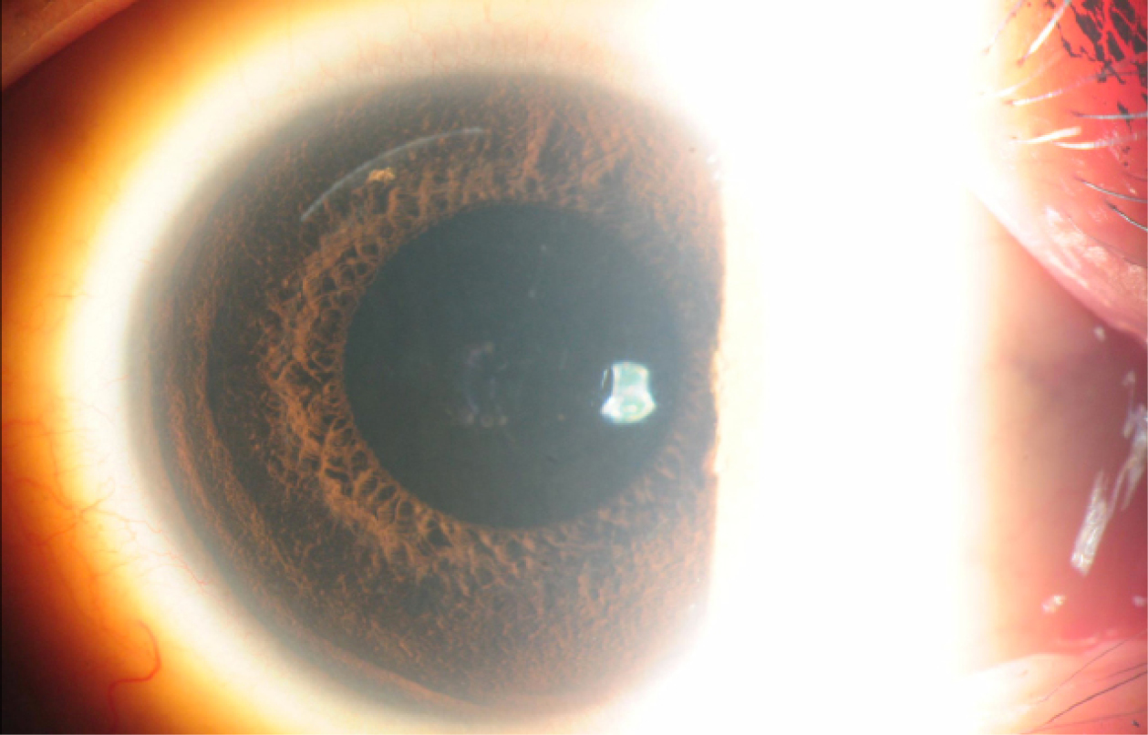
The increasing success of refractive surgery is based on technological advances that ensure safety and efficacy. Femtosecond lasers have been widely used to fashion a corneal flap in LASIK procedures, followed by corneal ablation using a separate excimer laser. The advantages of femto-LASIK have been well established compared with microkeratome-assisted LASIK, but what about an all-femto refractive surgical procedure?
Recently, Carl Zeiss Meditec introduced a novel form of a single-step, all-femtosecond laser refractive procedure, without the need of an excimer laser. This procedure is exclusively performed with the company’s VisuMax laser platform and is called refractive lenticule extraction or ReLEx. The laser dissects an aspheric lenticule of predetermined power within the corneal stroma to perform the predicted refractive correction. The first step of this procedure is the creation of the lenticule’s posterior surface, followed by the anterior surface dissection, which extends beyond the limit of the posterior cut and is opened to the epithelial surface through a circumferential ring cut at its periphery. The resultant flap (similar to a LASIK flap) is then lifted, allowing the lenticule’s removal.
Owing to technological advances, the ReLEx technique evolved into the SMILE or small-incisionlenticule extraction procedure. In the SMILE technique, the dissected lenticule is removed through a small incision—which is approximately one-eighth the size of the incision made during a LASIK flap cut—without creation of a lamellar flap.

In the SMILE technique, the dissected lenticule is removed through a small incision— which is approximately one-eighth the size of the incision made during a LASIK flap cut—without creation of a lamellar flap.
ADVANTAGES OF THE ALL-FEMTO TECHNIQUE
ReLEx has several advantages over standard laser refractive procedures like LASIK or PRK. In addition to a shorter procedural time, the suction pressure is minimal during the treatment. Studies report that ReLEx is precise and accurate in treating refractive errors and can be used to treat spherical myopic refractive errors up to -10.00 D and up to 5.00 D of astigmatism.1 The postoperative refractive outcomes are similar to that of LASIK. Furthermore, prospective studies indicate an insignificant regression rate in highly myopic eyes and fewer induced aberrations compared with LASIK.
Last year, the hyperopic treatment software became available. Recent studies point out the need to improve its predictability and effectiveness.2 Several conditions, including stromal hydration, laser fluence, and fixation loss, are very difficult to control and can influence the outcomes of excimer laser procedures. With all-femto procedures, the accuracy of the femtosecond laser is the only parameter that affects tissue removal and is not affected by environmental conditions.
SMILE
Concerning the flapless SMILE technique, the anterior stromal nerve plexus is damaged to a lesser extent and the additive excimer ablation is not performed, resulting in minimal dry eye symptoms and a faster recovery.3,4
As previously mentioned, SMILE is a flapless procedure, and a small side cut incision is created at an approximate depth of 100 µm to 120 μm in the cornea for the lenticule’s extraction. The inherent benefit of this option is the elimination of complications related to the LASIK flap, including irregular flap profiles, thin flaps, buttonholes, and free caps. Regarding corneal biomechanics, SMILE is also superior to conventional LASIK. When compared to the unoperated cornea, simulated corneal models demonstrate that flapless refractive surgery maintains the magnitude and distribution of stress patterns.5
Few complications have been reported in the literature. The loss of suction during the lamellar cuts may occur where the procedure can be aborted and performed at a later date.1,3 This complication can also occur with other femtosecond laser platforms. Another potential risk is the incomplete removal of the stromal lenticule, and this can be avoided by checking the lenticule under the microscope immediately after the removal.
If the patient requires an enhancement to achieve the target refraction, PRK or LASIK are feasible options. A thin-flap femto-LASIK procedure should also be considered, to perform the ablation in the corneal cap previously created by the SMILE procedure.
EXPANDING THE SURGICAL OPTIONS
Several animal studies propose that lenticule reimplantation after cryopreservation would be a way to reverse the effects of laser refractive surgery.6 These studies demonstrate that reimplantation of the lenticule restores the corneal stromal volume and curvature to its original state, achieving the preoperative refraction. This procedure also has benefits such as the autologous nature of the tissue, absence of sutures, and less wound-healing response.
This treatment modality may also be an option in presbyopic patients who may benefit from monovision or a stromal lenticule acting as a corneal inlay. In postrefractive surgery ectasia cases, lenticule reimplantation can also be combined with corneal collagen crosslinking. We were pleased to present the best paper of the session at ASCRS this year for the first description of a combined SMILE and crosslinking technique, where riboflavin was introduced into the pocket.7
CONCLUSIONS
ReLEx is one of the most significant developments in corneal refractive surgery since the introduction of LASIK. The new SMILE technique combines the benefits of a small incision with the other advantages over excimer laser procedures. In the future, precise and accurate treatment for hyperopia and other surgical modalities will be available.


