
Many guys are drawn toward electronics. An admitted technology geek, I have always been excited about the different modalities we ophthalmologists have to evaluate the ocular structures. For quite a while, I was excited about the concept of widefield photography, particularly that which could be performed quickly without dilating the patient. Until recently, I was frustrated with the ultra-widefield imaging devices offered by Optos—the original model was too big for my startup space, and the newer Daytona model did not have the chin rests and headrests my older patients would require for good fixation. Enter the latest California model.
The Optos California offers the best of both worlds: a small footprint and the ability for either the patient or technician to adjust the head position to allow for the best images. In practice, it’s been outstanding. Patients of all ages have been able to use it, and image acquisition takes about a second to perform. Instantly, the fundus photos are available on all of my WiFi-enabled computers. Many would say, “Why do you need this? You’re an anterior segment guy.” To them, I would reply that this device is valuable for anterior segment people for multiple reasons, as outlined below.
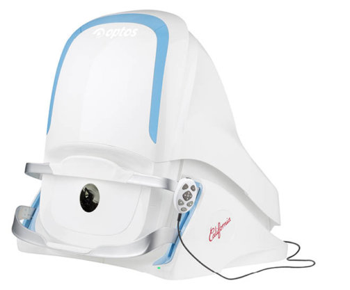
The Optos California.
PATIENT EXAMINATION
To begin, patients can sometimes be difficult to examine. Some are photosensitive, some refuse dilating drops, and others simply will not cooperate. Let’s face it, these were probably some of the patients who convinced us not to specialize in retina. Although I still perform dilated examinations on all new patients, I am much more comfortable having the Optos image beforehand to confirm my findings. In some cases, it can help me locate peripheral pathology such as a nevus before I even examine the patient.
PRACTICE EFFICIENCY
Because image acquisition is so quick, I have forgone dilating any postop month 1 cataract or YAG laser patients who are asymptomatic. Instead, a fundus photo is taken and, if normal, the patient can avoid dilation. This not only makes the patients much happier but it also shortens their visit time by nearly 30 minutes. It minimizes overcrowding in the waiting room as well.
PATIENT SATISFACTION
Although I mentioned patient satisfaction with no dilation, there are still several other ways in which this technology improves patient satisfaction. Many times, patients will come in with a history of retinal tear or a peripheral choroidal nevus, but they’ve never been able to appreciate what anything actually looks like. Demonstrating the tear with surrounding laser demarcation spots not only allows for better patient education but also significantly increases the “wow” factor.
PATIENT EDUCATION
The Optos California includes software to demonstrate pathology such as cataracts and changes with monofocal/multifocal lenses overlaying the patient’s own retina to provide accurate representations of cataract surgery.
FINANCIAL ASPECT
I mention this last because most practices already have fundus cameras. Of course, having widefield peripheral ability does allow for more pathology to be captured, and photos can still be performed without dilation. Nevertheless, for me, this is the least important reason to acquire the device.
CONCLUSION
There are clearly many more functions to this device, although many of them may be of greater value to retina specialists. That said, after only a month with this device, I can easily state that this is an outstanding device for any anterior segment surgeon.


