


SPEAKERS
Shamik Bafna, MD
• Cleveland Eye Clinic, Brecksville, OH

Ashley Brissette, MD, MSc, FRCSC
• Weill Cornell Medicine, New York, NY

Steve Charles, MD, FACS, FICS
• Charles Retina Institute, Germantown, TN

Steven D. Vold, MD
• Vold Vision, Fayetteville, AR
*Surgeons are paid Alcon speakers and consultants
Using the NGENUITY 3D Visualization System (Alcon) is like “watching a movie on steroids,” says Steven D. Vold, MD, Fayetteville, Arkansas.
Dr. Vold, who has been using ocular-free 3D visualization in the OR for more than 10 years, says the NGENUITY system has advanced well beyond early versions of this technology.
“The quality of visualization, which is so important for microinvasive glaucoma surgery (MIGS) and complex cataract surgery, is unbelievable with NGENUITY,” he says. “We’re seeing details that we never saw before.”
Dr. Vold; Shamik Bafna, MD; Ashley Brissette, MD, MSc, FRCSC; and retina specialist Steve Charles, MD, FACS, FICS, shared their insights on 3D visualization with the NGENUITY system at a special session during the 2019 meeting of the American Society of Cataract and Refractive Surgery. Here, we break down the features and functionality they appreciate.
Inside the System
The NGENUITY System features a 3D high dynamic range (HDR) camera, an ultra high-speed image processor, and a surgical 55-inch, 3D 4K organic light-emitting diode (OLED) surgical display that the team views with passive, circularly polarized 3D eyeglasses.
The HDR camera, which replaces the oculars and attaches to most surgical microscopes, produces true stereoscopic 3D imaging with automatic gain control and no light saturation. It requires no alignment, focus, or synchronization.
“The HDR camera has two 1080p chips,” Dr. Charles explains. “‘Why not 4K?’ you may ask. The reason is this: the optical system’s point spread function must match the pixel size, which is something cataract surgeons understand very well. If pixel size doesn’t match the optics, there would be zero advantage.”
The 3D HDR technology optimizes surgical images in real time. It balances light to mitigate glare and shadows to ensure that details and colors are highlighted effectively.
“Dynamic range is the difference between the least bright and the brightest areas in the image that can be displayed,” Dr. Charles says. “While the system does not produce images at the same dynamic range as our eyes, it has the best HDR sensors available. As shown in Figure 1, by averaging two frames, the image processor optimizes the view of the river as well as the brightest highlights, as it compresses the dynamic range into the visible range for us.”

Figure 1. The image processor compresses the dynamic range into the visible range for a clear view of color, contrast, and detail.
My First NGENUITY Case
by Ashley Brissette, MD, MSc, FRCSC
My first case using the NGENUITY 3D Visualization System was a 53-year-old man with a large, progressive pterygium of the left eye that was inducing irregular astigmatism and blurry vision and was cosmetically bothersome. My surgical plan was pterygium excision with mitomycin C and amniotic membrane grafting.
This case was ideal for evaluating the NGENUITY System for overall visualization, ergonomics (I expected the surgery would take at least 45 minutes), and for its value as a teaching tool, because a resident would be present and possibly participate in the case.
Any concerns I had about getting used to this new way of visualizing my surgery were quickly dispelled. It was actually quite easy to just sit down and start operating.
One remarkable feature that I recognized in this case—something that is really important for anterior segment surgery—was the depth of field and the peripheral acuity that I didn’t know I was missing before. Everything from the center all the way to the last pixel at the periphery of my case was in focus. As I was operating, I was seeing everything in HD 4K quality. I didn’t have to adjust my microscope or move the patient’s head or the microscope during the surgery.
Another great advantage of the NGENUITY system was that my resident and I were able to switch back and forth during the surgery without needing to adjust the microscope height or the oculars. Everything was in focus for both of us.
Not only were my resident and I engaged in this case, but everyone in the OR was involved. My scrub nurse could anticipate what I would need next, because she was watching along with us. She was more efficient because she was involved in the case.
—Ashley Brissette, MD, MSc, FRCSC
The ultra high-speed image processor simultaneously streams 3D and 2D to multiple display types and formats, and it produces real-time compressed 3D video files. It can record and save more than 100 hours of 3D video on its internal hard drive.
“The video recording capabilities are wonderful,” Dr. Brissette says. “As a newer surgeon, I’m building my repertoire of cases, and the quality of the images that can be recorded is great.”
Dr. Bafna notes, “Thanks to the powerful ultra high-speed image processor, we can see our maneuvers immediately in real time. Depth of field, magnification, and image resolution are fantastic.”
The 55-inch 4K OLED display provides an immersive view with exceptional contrast sensitivity.
“Why do we have a 4K display when we don’t have 4K camera chips? Is this somehow a disadvantage?” Dr. Charles asks. “Absolutely not. It’s 4K because the huge 55-inch screen with a 16x9 aspect ratio gives us four corners surrounding the circular surgical image for displaying various modes of the machines we’re using—in my case, the Constellation Vision System (Alcon)—so that I can view intraocular pressure, cutting rates, and various other data. In that sense, the NGENUITY system is somewhat like a heads-up display in an airplane cockpit, but the big advantage is improved visualization. That’s what this is all about.”
As for the OLED feature, Dr. Charles notes, “The OLED allows the surgeon to use high-acuity photopic vision rather than mesopic vision to view fine spatial details. When something is black, it’s black, which is not true of active-matrix LCDs.”
Visualization Benefits
The NGENUITY 3D Visualization System delivers an extended depth of field up to five times that of a traditional analog microscope (Figure 2).1 “This has been calculated. It’s been measured. It’s supportable,” Dr. Charles says. “Frankly, the FDA wouldn’t let us say it unless it were true. That’s a tremendously big deal.”

Figure 2. The NGENUITY 3D Visualization System delivers up to 5 times extended depth of field compared to a traditional analog microscope.
According to Dr. Bafna, “Once you’ve optimized your focus, the extended depth of field enables you to see from the cornea all the way down to the posterior capsule.”
The NGENUITY System also outperforms the analog microscope in terms of magnification, with up to a 48% increase,1 and edge-to-edge image clarity.
“One of the drawbacks with most analog microscopes is that the center of the image is clear, but the image tends to degrade at the periphery,” Dr. Bafna says. “That is a non-issue with the NGENUITY System. We now have vibrant clarity from edge to edge. Image resolution is much superior to the traditional analog microscope. These benefits are particularly important for surgeons performing MIGS.”
Education, Collaboration, Efficiency
In addition to presenting a crisp, clear image to the surgeon, the NGENUITY 3D Visualization System offers a 3D view of the surgical field to everyone in the OR, facilitating education, training, and collaboration, and fostering efficiency (Figure 3).
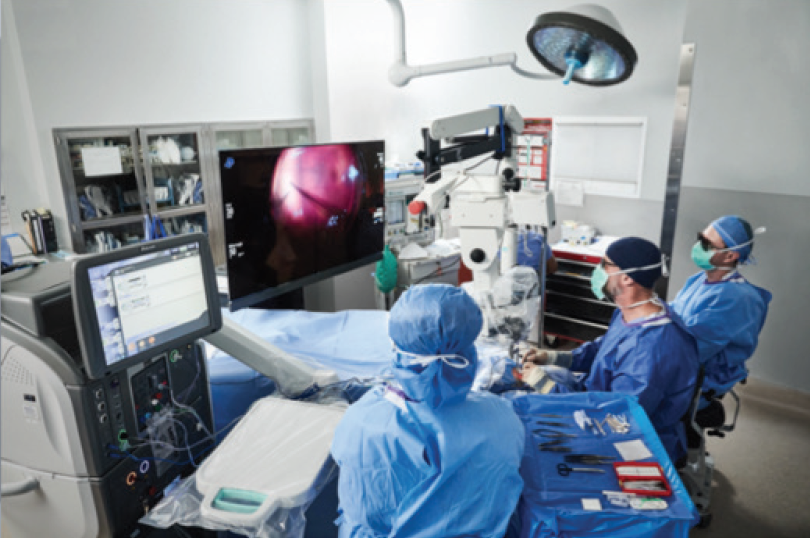
Figure 3. Whether the goal is to educate, train, or collaborate, this system provides an unparalleled view of real-time surgical information.
“The NGENUITY system is a phenomenal teaching tool,” Dr. Vold says. “It changes the game entirely.”
Dr. Brissette agrees. “The residents love it,” she says, “because they can be so much more engaged in the cases.
“An unexpected effect of NGENUITY in our OR is the team environment it creates,” Dr. Brissette continues. “Scrub nurses, circulators, even anesthesiologists, are more engaged in cases and better able to anticipate what I might need next. It has improved efficiency for our team.”
According to Dr. Charles, “The amount of engagement among everyone in the OR is difficult to appreciate until you see it. Everyone sees exactly what the surgeon sees. It is incredible. It’s tremendous for team teaching and team coordination. In the cockpit, we call it crew resource management. I really like that aspect of NGENUITY.”
Ergonomic Advantage
In addition to exceptional visualization, the ocular-free design of the NGENUITY System is intended to improve posture and may reduce fatigue.2
Dr. Brissette has a personal interest in the ergonomics of ophthalmic surgery and has researched the risk factors and consequences of poor posture in the OR.
“Rates of back and neck discomfort are extremely high among surgeons,” she says. “The higher our surgical volumes, the more we operate—even the number of patients we examine at the microscope in clinic—all of this takes a toll on our bodies over time. More than 70% of ophthalmologists have musculoskeletal symptoms, and up to 9% of surgeons have had to stop operating at some point in their careers due to pain or spine injury because of discomfort from their use of the OR microscope.3,4 The more I talk to my colleagues, the more I realize how important a physician’s well-being is to career longevity.”
Maximizing Visualization With the NGENUITY System
Dr. Charles offers the following pearls to maximize your viewing experience with the NGENUITY System:
- Set the camera’s aperture at 30% or one-third from the left side. If you set it in the middle, you won’t have the depth-of-field advantage.
- White-balance with the microscope light source and the room lights off. Do that properly and the color rendition is terrific.
- Position the display 4 ft away.
- Fill the screen vertically; otherwise, there’s no magnification advantage. Focus on what you’re working on at the highest magnification.
NGENUITY WITH DR. CHARLES
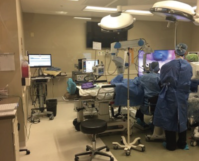
Figure 4. Shamik Bafna, MD, operates using the NGENUITY 3D Visualization System.
At 6’ 4”, Dr. Vold knows he’s at risk for back and neck problems as an ophthalmic surgeon. “Ergonomics is a big deal for me,” he says. “To be able to sit up straight while performing surgery or assisting my fellow in surgery, as opposed to being hunched over a microscope for an entire day, is truly a benefit. Heads-up surgery allows freedom of movement while retaining access to oculars at all times. It’s amazing how much better my posture is. Over the course of 10 to 20 years, that can make a big difference in my orthopedic and neurologic status.”
Conclusion: You’ll Never Want to Go Back
The NGENUITY 3D Visualization System is a game-changer in ophthalmic ORs, providing surgeons—and everyone else in the room—with a high-definition, highly magnified, 3D view of the surgical field and their operations in real time.
“The three main features that stand out for me about this system are, first, the visualization—you don’t know what you’re missing until you see it— followed by the education opportunities and the ergonomic advantages,” Dr. Brissette says.
Dr. Bafna notes, “Being able to visualize our surgeries so much better than before increases our confidence in what we can achieve.”
Dr. Vold recommends easing into ocular-free visualization by using it to perform some of the more straightforward cataract surgeries. “Once you get used to it, it becomes second nature,” he says. “Now, I really wouldn’t want to go back to using a microscope again.”
Dr. Charles has been using the NGENUITY System almost exclusively since 2016. “I use it on all of my cases except the few that I need to perform in the hospital. Having to go back to the microscope every now and then reminds me why I like NGENUITY.”
Participants are paid consultants to Alcon.
NGENUITY and CONSTELLATION are trademarks of Alcon. All other brand/product names are the trademarks of their respective owners.
© 2019 Alcon Inc. 08/19 US-NGU-1900071
1. Alcon data on file. Alcon Laboratories, Inc. December 2017.
2. Eckardt C, Paulo EB. Heads-up surgery for vitreoretinal procedures: an experimental and clinical study. Retina. 2016;36(1):137-147.
3. Kitzmann AS, Fethke NB, Baratz KH, et al. A survey study of musculoskeletal disorders among eye care physicians compared with family medicine physicians. Ophthalmology. 2012;119(2);213-220.
4. Sivak-Callcott JA, Diaz SR, Ducatman AM, et al. A survey study of occupational pain and injury in ophthalmic plastic surgeons. Ophthalmic Plast Reconstr Surg. 2011;27(1):28-32.
IMPORTANT PRODUCT INFORMATION FOR NGENUITY® 3D VISUALIZATION SYSTEM FOR THE DIGITALLY ASSISTED VITREORETINAL SURGERY PLATFORM
IMPORTANT PRODUCT INFORMATION
CAUTION: Federal (USA) law restricts this device to sale by, or on the order of, a physician.
INDICATION: The NGenuity® 3D Visualization System consists of a 3D stereoscopic, high-definition digital video camera and workstation to provide magnified stereoscopic images of objects during micro-surgery. It acts as an adjunct to the surgical microscope during surgery displaying real-time images or images from recordings.
WARNINGS: The system is not suitable for use in the presence of flammable anesthetics mixture with air or oxygen. There are no known contraindications for use of this device.
PRECAUTIONS: Do not touch any system component and the patient at the same time during a procedure to prevent electric shock. When operating in 3D, to ensure optimal image quality, use only approved passive‐polarized glasses. Use of polarized prescription glasses will cause the 3D effect to be distorted. In case of emergency, keep the microscope oculars and mounting accessories in the cart top drawer. If there are any concerns regarding the continued safe use of the Ngenuity® 3D Visualization System, consider returning to using the microscope oculars.
ATTENTION: Refer to the User Manual for a complete list of appropriate uses, warnings and precautions.


