


It is well known that the earlier glaucoma is detected, the better the patient’s prognosis will be; however, it is not always easy to determine the onset of glaucoma with the use of visual field testing or optical coherence tomography (OCT) alone. Using these diagnostics in conjunction with electrophysiologic testing of the retina and neurovisual pathways may provide a more complete picture of the patient’s condition.
Visual evoked potential (VEP) and pattern electroretinography (pERG) tests can be invaluable in helping clinicians detect the onset of disease. Together with other functional and structural tests, the NOVA Vision Testing System (Diopsys), which features both VEP and ERG objective functional tests, has enhanced my ability to detect disease and intervene at a point when there is still time to halt irreparable damage.
BREAKING IT DOWN: VEP AND ERG
In general, VEP involves measuring the electrical activity of the patient’s visual pathway, from the retina to the visual cortex (Figure 1). The signal’s amplitude measures how much information is reaching the visual cortex and will be larger or smaller depending on the disease state. Latency of the signal measures the length of time it takes to travel from the retina to the visual cortex. Anything that impedes conduction of the electrical signal, such as optic neuritis or multiple sclerosis—both issues that affect the myelin sheath—can cause increased latencies.
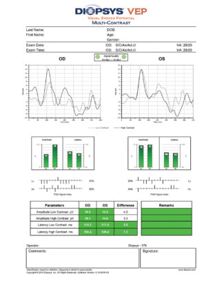
A sample normal Diopsys VEP multicontrast report.
Patients are tested at low contrast (15%) and at high contrast (85%). The high contrast measures the function of the parvocellular pathways, which is an early indicator of central vision loss. The low contrast measures the function of the magnocellular pathways, which may be an early indicator of glaucoma.
pERG measures the function of ganglion cells in the retina, which is useful in providing information for diseases that have an expected pattern of macular degeneration. pERG has two types of stimuli, the first being contrast sensitivity using a high and a low contrast stimulus, which is designed to provide data to aid in the diagnosis of diseases that affect the retina in a diffuse pattern, such as chronic open-angle glaucoma and diabetic retinopathy. Second, the concentric stimulus fields protocol tests the retina at 24° and 16° and is intended to be used to aid in the diagnosis of diseases that affect the retina in a specific topographic pattern, such as age-related macular degeneration and diabetic macular edema, and toxic maculopathies. These types of diseases usually affect the central and paracentral area of the macula.
VEP and ERG are objective, functional tests. As they do not rely on subjective, manual responses from the patient, the results gathered have strong credibility. This is one reason why these tests are also useful for malingerers, non-verbal patients, children, or those otherwise unable to take an active role in testing. VEP and ERG can also be used to detect functional changes at very early stages of disease. Once there is structural damage to cells in the retina, it is permanent and irreversible. By catching disease prior to cell death, it is possible to restore distressed cells and possibly reverse disease progression.

Handheld mini-Ganzfeld for Diopsys full-field ERG vision testing.
UPCOMING TECHNOLOGY
I have been involved in the beta testing of the Diopsys mini-Ganzfeld device (Figure 2), which measures the electrical response of the visual system via full-field flash ERG testing. This test is designed to allow retina specialists to look at rod-cone dystrophies and to help cataract surgeons attribute percentages of vision loss to either the cataract or an underlying maculopathy, such as age-related macular degeneration and/or an epiretinal membrane.
Just like VEP, the data are delivered in a graphic presentation that represents quantitative measurements. Our experience so far shows the tests to be repeatable with cataract patients and demonstrates clear functional vision loss due to age-related macular degeneration and diabetic retinopathy. We believe that this new full-field ERG module with the mini-Ganzfield device will be useful in diagnosis, decision-making regarding patient therapies, and IOL selection in cataract patients.
A COMPLETE CARE PACKAGE
Early detection of glaucoma is paramount to establishing an improved prognosis. Structural damage revealed by an OCT test is permanent and irreversible. However, utilizing visual electrophysiology along with other standard tests in my practice has allowed me to detect suffering ganglion cells in time to treat patients and potentially reverse the disease if caught early enough.
I do not perform VEP and pattern ERG alone. OCT measures structure, and there is great value in that information. I continue to perform visual field tests and OCT images of the optic nerve in order to establish the whole picture of the patient. I use electrophysiology for patients with ocular hypertension, suspected glaucoma, established glaucoma, normal-tension glaucoma, most maculopathies, any retinopathies, hydroxychloroquine or other drug toxicity, and suspected malingerers.
CONCLUSION
Visual electrophysiology is certainly not a substitute for other forms of testing, but it is a welcome addition to my practice, adding to the global analysis for most of these conditions. Most critically, it allows me to measure and determine concerns and issues earlier. In fact, a recent study showed that pattern ERG signals were able to show ganglion cell loss an average of 8 years earlier than with OCT.1
As the tests measure the functionality of the retinal ganglion cell complex, I can also determine the efficacy of treatment, which allows me to ascertain if therapy is working or needs to be modified. Being able to catch these problems early when the ganglion cells are suffering and there is still a possibility to prevent further damage can make a monumental difference. Visual field, OCT, and electrophysiology combined provide a complete package and should be considered the standard of care.
1. Banitt MR, Ventura LM, Feuer WJ, et al. Progressive loss of retinal ganglion cell function precedes structural loss by several years in glaucoma suspects. Invest Ophthalmol Vis Sci. 2013;(54):2346-2352.