supported by


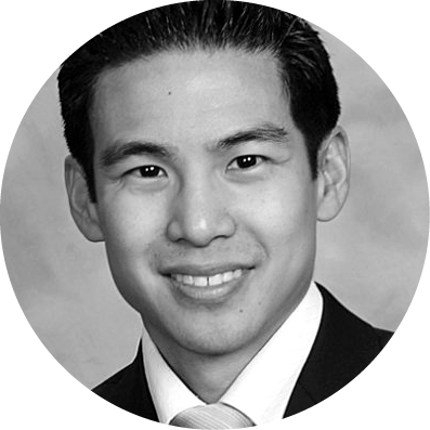
Matthew Feng, MD
Dr. Feng is a corneal and refractive anterior segment surgeon at Price Vision Group in Indianapolis, Indiana.
Please share with us your background.
Home is Pittsburgh, Pennsylvania, where my parents still reside after emigrating from Taiwan. I am a product of the western Pennsylvania work ethic, the Pittsburgh public school system, and the immigrant narrative. My graduation from Taylor Allderdice High School (go Dragons!) was an especially proud day for my father, for when I was introduced as co-valedictorian, the superintendent accidentally called my father’s name, not mine. It was a fitting and symbolic affirmation of his sacrifice and my mother’s.
My earliest passions were architecture and classical piano. I played at Carnegie Hall three times through the American Music Scholarship Association and nearly pursued a career in music. Academically, I was blessed to be selected as a Presidential Scholar by the Department of Education and an inaugural Millennium Scholar by the Bill and Melinda Gates Foundation. I graduated magna cum laude from Harvard with a degree in biochemical sciences, then worked at the Children’s Hospital Boston and Dana-Farber Cancer Institute before accepting a Dean’s Merit Scholarship to return home and study at the University of Pittsburgh School of Medicine.
What drew you to ophthalmology and, specifically, to your field of interest?
I was a third-year medical student intent on orthopedic surgery, specifically of the hand, when I began a 1-week ophthalmology rotation. Truth be told, it was just a 3-day rotation, as I had scheduled the experience the week of Thanksgiving—so sure that I was uninterested in ophthalmology. Instead, the first day was a whirlwind of epiphanies. By the time I watched that night’s on-call resident relieve an acute angle-closure attack via laser iridotomy, I wanted those holiday days back.
I love art and science, clinical medicine and microsurgery, and optics and lasers. Once I realized that ophthalmology represented the intersection of all these elements, I knew a sea change was coming. Jake Waxman, MD, Associate Professor of Ophthalmology and Residency Director, was an invaluable guide as I switched specialty interests. Things could not have worked out better in the end, as my wife, Brenna (emergency medicine), and I each wound up in Arizona for our respective residencies.
There, in the dry desert air, I discovered a predilection for the anterior segment and, most importantly, discovered supportive faculty mentors who shared the same. I found particular satisfaction in treating reversible conditions ranging from simple refractive error to cloudy corneas to restore or even enhance sight. I enjoyed having a surgeon’s skill and attention to detail tangibly rewarded in letters or even lines of uncorrected vision. That said, it is even more exciting to have one’s diagnostic and surgical skills augmented by ever-expanding anterior segment technologies.
Please describe your current position.
I am an anterior segment surgeon in private practice. My patient mix is primarily cornea and external disease, cataract, glaucoma, and refractive. My time is split approximately two-thirds in a busy tertiary referral clinic and one-third operating. I perform Descemet membrane endothelial keratoplasty (DMEK), Descemet stripping automated endothelial keratoplasty (DSAEK), deep anterior lamellar keratoplasty (DALK) and penetrating keratoplasty with and without laser assistance, intrastromal corneal ring segments, CXL, keratoprosthesis, anterior segment reconstruction, traditional glaucoma surgery, MIGS, complex cataract surgery, pars plana vitrectomy with and without pars plana lensectomy, glued and scleral-sutured IOLs, premium IOLs, Visian ICL (STAAR Surgical), LASIK, PRK, pterygium excision, and more. Also, the practice trains one to two fellows annually, and it is a privilege to be an instructor.
Who are/were your mentors?
Hanna Wu Li, Professor of Piano and Piano Pedagogy at Carnegie Mellon University, was the first to teach me manual dexterity, pedal work, and discipline. Andy Eller, MD, Professor of Ophthalmology at the University of Pittsburgh, gave me a chance on his retina service as a medical student after I realized—almost too late—that I loved ophthalmology, not orthopedics.
At the University of Arizona, mentors were myriad. Department Head Joe Miller, MD, MPH, taught me optics and suturing; Bill Fishkind, MD, and Brian Hunter, MD, phacoemulsification and chopping techniques; Roxana Ursea, MD, cornea and anterior segment imaging; and Michael Belin, MD, corneal tomography and keratoplasty. Finally, I moved to Indiana, where I was fortunate to complete my corneal fellowship (really an anterior segment fellowship) with Barraquer Award recipient and endothelial keratoplasty pioneer Frank Price, Jr, MD.
What has been the most memorable experience of your career thus far?
It’s a close call. Case-wise, there was a young tearful mother with congenital cataracts and early-onset Fuchs dystrophy who came to me with bilateral aphakia, zero capsular support, and painful bullous keratopathy. After pars plana vitrectomy, secondary glued IOL placement, and then staged DMEK, she was 20/20 uncorrected and had her “life back.”
Then there was a Chinese national who had four failed keratoplasties overseas in his only functional eye. Cicatrization of his ocular surface was so bad that I had to perform complete ocular surface reconstruction with amniotic membrane and limbal stem cell autografts from his fellow eye just to identify his cornea and fornices. After staged cataract extraction, vitrectomy, and aphakic Boston keratoprosthesis placement, he saw his grandson’s face for the first time.
My most memorable day came 3 months out of fellowship, when Dr. Price called in sick on his operating day. His patients agreed to let me operate in his stead, and fortunately their trust was not misplaced. I performed more DMEKs in a day than I normally did at that time over several weeks. Throughout the day, I reminded myself how thankful I was for solid mentors and training. By that day’s successful end, I thought for the first time that maybe, just maybe, I could actually make it as a cornea surgeon.
What are some new technological advances that you have found particularly exciting? Which advances in the pipeline are you most enthusiastic or curious about?
Like many others in the United States who have given up on having a Schwind Amaris, I am excited to see what Alcon’s Contoura Vision can do. Topography-guided treatments should enhance our ability to correct regular refractive errors on the one hand and create new, sorely needed options for irregular corneas on the other.
What is the focus of some of your research?
DMEK has reached critical mass, where many corneal surgeons have now tried the technique. I am interested in further simplifying the technique to expand adoption, identifying donor and recipient risk factors to eliminate rare graft failures, and better predicting shifts in postoperative corneal power and cylinder via perioperative imaging to limit refractive surprises in triple cases. I am also studying rejection patterns associated with differing postoperative steroid regimens after DMEK and with differing techniques for DALK.
What is a typical day in your life? What keeps you busy, fulfilled, and passionate?
I rise early, drop off my still-somnolent children at preschool, and then care for as many patients as I can. Eating lunch is a plus. Most days, I can pick up the kids, enjoy dinner with the family to hear about everyone’s day, and then go a few rounds in the nightly tug of war that is putting the little humans to bed. Nevertheless, my talented and beautiful wife, true to her multitasking emergency medicine nature, is really the competent one who makes our family work. I love ophthalmology, and protecting family time allows me to recharge and return the next day 100%. If you ask again in a few years when Kira, 4, and Cameron, 2, are (hopefully) more self-sufficient, I might also credit some rediscovered hobbies such as piano, photography, and travel.
What advice can you offer to individuals who are just now choosing their career paths after finishing residency or fellowship?
Explore widely, and then pursue what excites you most from among your discoveries. Ask yourself, what am I doing and in what environment on days when I leap out of bed? On days when I crawl out of bed? Make sure that you love a subspecialty’s content and patient population, not just the charisma of that subspecialty mentor. Make especially sure that you love the bread-and-butter of your planned career. If you love it all, try to anticipate where the most future growth will be and position yourself to be there.
If anticipating proves difficult, try asking your patients. For me, I chose training that featured DMEK while it was highly controversial among surgeons because many patients had already decided for us. Fuchs patients who had DMEK in one eye and DSAEK in the other were telling others over social media that they needed DMEK and should travel cross-country if necessary to get it.
Finally, some practical advice. Purchase term life insurance (and create a will) once you have dependents or family members for whom you provide. Create a retirement plan and start saving if you have not already. Calculate how much principal you would need in the bank for those family members to reasonably live off the interest as a supplement to any income they generate themselves. That should be the value of the insurance policy. Once you have saved that amount, the insurance policy is no longer needed. Therefore, the policy’s term period can be calculated based on how long you anticipate it will take to save the aforementioned amount. To decrease premiums, one potential strategy involves two policies—eg, half the value with a 10-year term and the other half with a 20-year term.
Tell us about an innovative procedure you are performing or a new imaging/diagnostic tool that has improved your practice.
Intraoperative OCT arrived last year and is both fun and functional. It allows real-time visualization of depth during DALK procedures, graft orientation during endothelial keratoplasties in significantly cloudy corneas, vault heights during ICL placement, and more. At the time of this writing, the Zeiss system seems to have a slight advantage over Haag-Streit, with greater OCT capability and more seamless incorporation with the operating microscope. The Haag-Streit can infuse its intraoperative OCT image into both microscope oculars, however, and more software updates are planned. Full disclosure: I use the Haag-Streit system and have no financial interests in either company.
As far as procedures, intrascleral IOL fixation, so-called glued IOL, is a technique that I recommend anterior segment surgeons have in their toolkit for complex cataract or dislocated lens cases where capsular support is currently or expected to soon be inadequate. Certainly, there remains a role for scleral-sutured techniques, which I employ primarily to salvage the capsular bag via modified capsular tension rings or segments to permit placement of premium lenses in that space. However, in other situations, the glued IOL can reliably result in stable and well-centered placement of a posterior chamber monofocal or even multifocal three-piece lens prosthesis. Between this and familiarity with pars plana vitrectomy as recommended by David Chang, MD, and others, I have not had a restless night before cataract surgery in 3 years, and that is a gift I feel compelled to pay forward.


