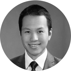
Ken Lin, MD, PhD
Dr. Lin is a Glaucoma and Cataract Surgeon at Anaheim Eye Institute and a Clinical Assistant Professor, Department of Ophthalmology, at the University of California, Irvine.
Please share with us your background.
I grew up in Taiwan and came to the United States during high school. Due to my limited English, I initially gravitated toward extracurricular activities that required little verbal communication. As such, I joined the school’s cross-country team. There are almost no strategies to discuss, and one just needs to run as fast and for as long as possible without stopping. I loved it, and the grit and physical fitness I acquired then still significantly affect my personal and professional life today.
I subsequently attended Stanford University. Being an undergraduate there in the early 2000s was an exciting time. Google was just starting, Netscape was still a popular browser, Apple had been resurrected with the success of the iMac, and genomics was the underlying theme of many biotech startups. Entrepreneurship was palpable in the air, and it is still the Zeitgeist there. This was also the birthplace of my interest in medicine, and my mentors there made my foray into biomedical research much easier. My parents rightfully pointed out that my research advisors were like my surrogate parents in Palo Alto. My lab colleagues from around the world bonded together like family members; this, I believe, was what enabled me to publish multiple peer-reviewed papers and patents as an undergraduate. After Stanford, I enrolled in Harvard Medical School’s MD-PhD Program, where I earned a PhD in biophysics. My doctoral thesis was in molecular imaging of pancreatic cancer. The imaging and microsurgical work I did as a graduate student was a major factor in my decision to pursue ophthalmology.
What drew you to ophthalmology and your field of interest?
Ophthalmology is arguably the most quantitative discipline in clinical medicine, and I have always been drawn to math and physical sciences. Part of my graduate thesis research involved implanting pancreatic ductal cells in a mouse pancreas. After the tumor grew, I developed protocols and techniques to perform in vivo imaging of the tumor cells and vasculature. That was my first foray into microsurgery and quickly became one of my favorite parts of research. I taught myself most of the physical optics at the time, and during the learning process I came across several retina journals. That was my first contact point with ophthalmology, and I quickly appreciated the eye as a perfect imaging system. Unlike the pancreas, which has the reputation of being one of the most difficult organs to image, the eyes are exposed and are much easier subjects for quantitative imaging. I was drawn to ophthalmology from a research standpoint.
In my third year of medical school, I had the opportunity to do an ophthalmology rotation during my surgery clerkship under the guidance of Kathryn Colby, MD, at the Massachusetts Eye and Ear Infirmary. Jessica Ciralsky, MD, who is now a cornea specialist at Cornell and a MillennialEYE Section Editor, was a first-year resident at the time. It was impossible to not enjoy ophthalmology after spending 3 weeks with these two incredible teachers. I liked the heavily exam-based clinic and loved the cataract and corneal transplant surgeries. My interest in both the clinical and research aspects of ophthalmology ultimately gravitated me toward the field.
Please describe your current position.
I have been a glaucoma specialist at a private practice since finishing fellowship a year ago, although comprehensive ophthalmology still comprises over two-thirds of my clinic. I also staff the glaucoma clinic at the Gavin Herbert Eye Institute at UC Irvine.
Who are/were your mentors?
John Cooke, MD, and Phyllis Gardner, MD, are my two Stanford mentors who inspired me to go into medicine. Throughout my ophthalmology residency, my department chair Roger Steinert, MD, was a great teacher. He treats residents like colleagues, spends a great deal of time mentoring, and constantly encourages innovation. During my chief residency year, he shared with me his rich experience in leadership, management, and industry. Finally, my glaucoma fellowship mentors, Sameh Mosaed, MD, and Anand Bhatt, MD, are two of the most gifted, caring surgeons I have ever known. It is impossible not to love glaucoma after learning from them. They taught me how to react under stress. I have come to appreciate that humans are like teabags—their true characters are not revealed until they are under hot water.
What is the most memorable experience of your career so far?
In residency, I performed extracapsular cataract extraction on a 78-year-old man with extensive health problems and brunescent cataracts in both eyes. His binocular BCVA was hand motion. Although he lived in a facility, his poor vision contributed to his depression and medication noncompliance. Under the microscope, his diabetic cataract appeared ready to pop out of the capsular bag at any moment. Despite my attending’s reminder to make a slightly bigger rhexis, I could not resist taking short-term comfort in keeping the rhexis “safe and small.” This, of course, made lens extraction much harder. Fortunately, after much struggle, the lens finally tilted and evacuated, and the case concluded uneventfully. The patient returned the next day with a UCVA of 20/40 in the operated eye. He was more bewildered than happy. His only full sentence at the visit was, “I felt like I just woke up from a long sleep.” “Welcome back, Captain America!” I congratulated him. We then set the surgery schedule for his other eye.
That was the last time I saw him. He never showed up to his postop appointments or second eye surgery. Eventually, we contacted the facility where he lived and were told he left. A few months later, I received a handwritten letter from the patient. He told me that his new vision has given him the strength to live again, so much so that he “escaped” from his facility and set out to reunite with his daughter. He told me not to worry about his other eye because “one good eye is enough for one happy soul.”
Which new technological advances do you find exciting?
I am exited about some of the surgical and glaucoma treatments emerging from the pipeline. On the medical side, latanoprostene bunod (LBN) has shown great potential to be a new ocular hypotensive agent. It is a prostaglandin receptor agonist and, structurally, is latanoprost tethered to a nitric oxide-donating moiety. Since the late 90s, nitric oxide has been known as a key mediator of vasodilation, and its reduction is a major cause of pathology in most cardiovascular and prothrombotic diseases. Nitric oxide synthase in vascular endothelial cells secretes nitric oxide, which diffuses to the neighboring vascular smooth muscle cells to effect relaxation through a cyclic GMP-dependent mechanism. Like smooth muscles, human trabecular cells also exhibit contractile property, and a growing body of evidence suggests that nitric oxide can reduce trabecular resistance.
Elevated levels of an endogenous inhibitor of nitric oxide have also been found in the aqueous of patients with primary open-angle glaucoma. It has been theorized that nitric oxide may also reduce episcleral pressure, but the preclinical data have been equivocal so far. Currently, phase 3 trials of LBN, such as APOLLO and LUNAR, have shown LBN having significantly greater IOP reduction than but a similar safety profile to latanoprost and timolol. Given my previous research background in nitric oxide, I naturally feel very excited about this possible new medical treatment for glaucoma.
I look forward to more results on MIGS stent devices such as CyPass (Transcend Medical), Hydrus (Ivantis), and Xen (Allergan). Recently FDA-approved, CyPass opens a conduit to the surpachoroidal space; the stent is made out of polyimide material, which is often used in IOL haptics. Hydrus is an intracanalicular scaffold made out of an alloy called nitinol. Nitniol has shape memory and is widely used in cardiovascular and orthopedic implants. When inserted into the Schlemm canal, Hydrus stents the canal along its 3-clock-hour length and theoretically allows for more access to the collector channels. Xen is a microstent that drains to the subconjunctival space. Made out of porcine gelatin, Xen is flexible and noninflammatory. Once hydrated, it swells and locks itself within the scleral tissue to minimize movement post implantation.
What is the focus of some of your research?
Part of my research focuses on developing quantitative imaging techniques to identify areas of high and low flow in the intrascleral portion of the conventional outflow pathway. Such information may help target MIGS devices to areas with high flow to maximize IOP reduction. Another aspect of my research is developing smartphone-based software to improve patients’ medication compliance.
What is a typical day in your life? What keeps you busy, fulfilled, and passionate?
I operate two afternoons a week and maintain a busy clinic the rest of the week. I exercise daily, jogging at least 1 or 2 miles and swimming with my kids. Finding balance between work and family is not always easy, but being efficient with one’s time helps. I often remind myself that if a task takes 5 minutes to do, I shall complete it in no more than so. I am definitely not compliant with this principle at all times, but when I am, a surprising amount of free time surfaces on my schedule. I find pleasure in everything I do and try to see the world through a positive lens—positive as in optimistic and not hyperopia-correcting. This mentality helps me achieve a sustained feeling of fulfillment and happiness.
Most patients look forward to their appointments with ophthalmologists with a certain degree of anticipation. The check-in, testing, and workup are akin to drumrolls that all lead up to the finale of meeting the surgeon. On the other hand, our days as surgeons are comprised almost entirely of those finales. As such, what we regard as routine may be perceived by patients as anything but. Some wait weeks to ask their questions during that finale, but, on our schedule sheet, that is but one of the forty or so encounters for the day. This dichotomy between patients’ and doctors’ states of mind is a good reminder that the least I can do for my patients is listen to them.
What advice can you offer to individuals who are choosing their career paths after finishing residency or fellowship?
It helps to start looking for job openings early, especially if you already have a geographic preference. Some practices may not have openings at the moment, but they may anticipate the need in 2 years due to office expansion or senior partners retiring. As such, one can start the search process prior to starting fellowship or PGY4. Local pharmaceutical sales representatives are often knowledgeable of potential openings in their areas before the positions are publicly advertised.
Most trainees know the salient differences between academic and private practice careers. In my experience working with residents, however, these differences are often not distinguishing enough to precipitate a clear preference. In fact, the distinctions between these two career paths have blurred over the years. Numerous landmark clinical papers have come from private practice investigators, and many academicians are now thought leaders who work with industry to innovate from bench to bedside. There are aspects of teaching, research, administrative, and clinical work that we all enjoy. For the most part, young ophthalmologists can explore and incorporate those aspects in either setting. I encourage trainees to not pigeonhole themselves into one category too early. Make a list of what you value most in life over the next 10 years and prioritize each entry. Such a thought process often indirectly helps reveal a career path.
In addition, faculty members are excellent mentors. They have gone through the process, many have raised a family, some have switched practice settings, and surely each has his or her personal story to share. Last, keep in mind that each locale is unique. The advice you receive about starting a career in one county in California may not be entirely applicable to other states or even other parts of California.
Tell us about a new procedure or diagnostic tool that has improved your practice.
Intraoperative aberrometry with ORA (Alcon) is an integral part of my astigmatism management during cataract surgery, especially at a time when posterior corneal astigmatism is receiving more attention. Like neutrinos or other subatomic particles, posterior corneal astigmatism has an elusive air around it—we know it exists, but its direct measurement is difficult, and we often rely on several indirect data to help define it. While the Graham Barrett formula generally gives accurate power and axis information, ORA provides another data point that quantitatively accounts for posterior corneal astigmatism. It also helps refine toric IOL alignment. However, knowing when to ignore ORA data is just as important as knowing how to incorporate it. A deep-set orbit with a tight lid speculum, corneal edema, retinal pathology, air bubble, and stromal opacity are just some of the many factors that can alter ORA readings.


