


In medical training, practice makes progress. We cannot master a surgical technique until we try … and try again. Thus, the use of surgical training devices is becoming more popular and more important with time.
The Idrees Eye Surgery Practice Head (Gulden Ophthalmics) is an innovative device for surgical training. This product is designed to hold pig, sheep, or cadaver eyes, enabling trainees to hone their surgical skills and build confidence without posing any danger to human eyes. The LED illumination system provides an excellent red reflex for a realistic surgical experience.
This article describes the need I saw to create such a device, the path to its development, and its ability to maximize skills transfer from training to a practice setting.
Video | The Idrees Eye Surgery Practice Head.
DEVICE DEVELOPMENT
My keen interest in training young eye surgeons was the driving force behind the development of the Idrees Eye Surgery Practice Head. I conceived the idea while working in a charity hospital in Pakistan and training junior doctors. The charity hospital was not equipped with any facility or device to teach eye surgery. I could see that most of the junior doctors were eager to learn, and, at the same time, I was keen on teaching them to restore sight to the less-privileged patients in the community.

Figure 1 | REDO Eye Hospital, Rawalpindi, in Pakistan.
I started searching for a satisfactory device to train these doctors in the charity setup. However, I could not find anything that would allow the transfer of surgical skills in a safe and successful manner. I was looking for something that could hold an animal eye firmly in the desired position, provide a bright fundus glow, keep the eye well pressurized during the procedure, have a drain for irrigation fluid, and provide comfortable positioning for the surgeon while working.
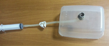
Figure 2 | The basic prototype.
Realizing this did not exist, I started brainstorming a new device. Once I conceived the initial idea, I purchased a few dollars’ worth of solid metal and had it customized to feature an eye-holding cup. I fitted a tube with a lock and connected that to a 20-cc syringe (Figure 2). I then got a plastic container, fitted the device model onto that, and tried to teach the trainees with a sheep eye. It worked well, but I found there was a need to fit an illumination device at the base of the eye-holding cup to provide the fundus glow.
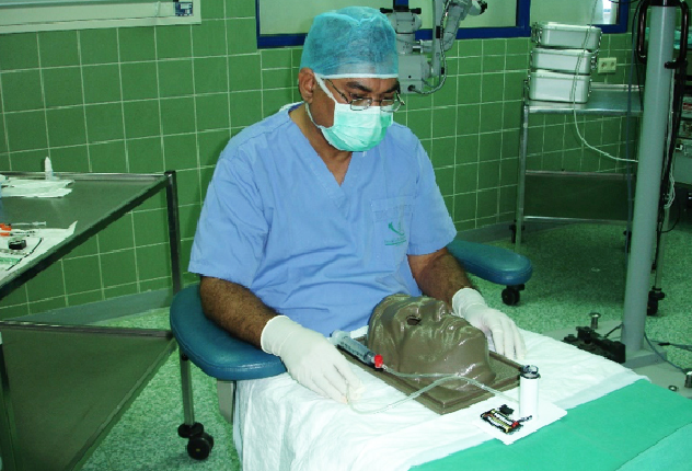
Figure 3 | The first prototype with LED illumination and the vacuum device.
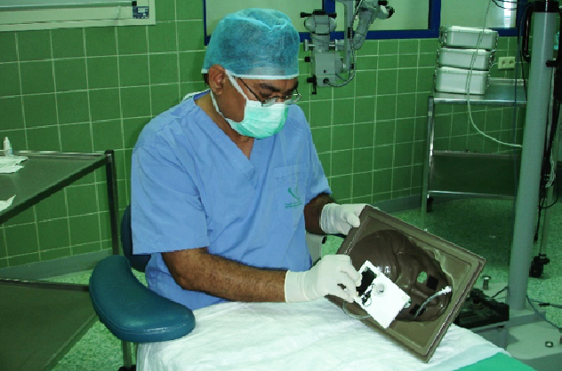
Figure 4 | Assembling the first prototype for final testing.
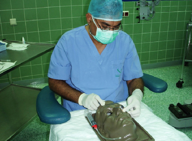
Figure 5 | First prototype with a sheep eye in place (left side).
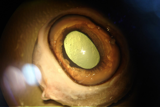
Figure 6 | Bright fundus glow of the sheep eye under the microscope.
In 2014, I traveled to Boston, Massachusetts, to present two papers at the annual meeting of the American Society of Cataract and Refractive Surgery. While there, I met Tom Kockley of Gulden Ophthalmics and discussed with him this training device. Following that conversation, a prototype of the practice head with an LED light at the base of the eye-holding cup (Figures 3 through 5) was created. The prototype worked well with the bright fundus glow (Figure 6), so we moved to the final version of Idrees Eye Surgery Practice Head (Figure 7).
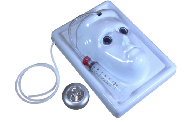
Figure 7 | The final product.
APPLICATIONS
Teaching eye surgery to ophthalmology residents is the main use of this device. This practice head provides stress-free training for young ophthalmologists beginning their surgical careers. If the training is under supervision of a more experienced surgeon, the trainee’s learning can be further facilitated by his or her guidance. The device can also be useful in wet lab sessions. In my experience, training on this device usually translates to efficient surgical skill.
As we work to master new techniques, technology continues to advance. The Idrees Eye Surgery Practice Head can help surgeons keep up with the innovative products and approaches that arise. The surgical procedures and techniques that can be practiced with this device include:
- Manual small-incision cataract surgery
- Phacoemulsification
- Laser refractive corneal surgery (PRK and LASIK)
- Peripheral corneal incisions for astigmatism correction
- Penetrating keratoplasty
- Lamellar keratoplasty
- Corneal debridement
- Keratoprosthesis
- Corneal glue application for sealing corneal perforations
- Corneal collagen crosslinking
- Trabeculectomy
- Scleral suturing for squint surgery
- Intravitreal injections
- Vitreoretinal surgery
- Endolaser treatment of the ciliary body and retina
- Surgical procedures on the iris
- Repair of perforating corneoscleral injuries
THE SETUP
Setting up the Idrees Eye Surgery Practice Head is fairly simple. First, put on gloves. Next, position the practice head under the microscope. Then place a sheep, pig, or cadaver eye in the eye-holding cup, apply vacuum, and adjust the pressure in the eye for the required procedure. Lock the vacuum. Place the LED light under the device head if a fundus glow is needed. The level of background illumination can be controlled by placing the LED light at the appropriate place. Last, apply the drape, adjust the irrigation fluid-collecting tube, and focus the microscope. You can then proceed with the planned procedure.
Once the surgical session ends, remove the drape, release the vacuum, remove the eye, and dispose of any fluid collected in the container. Switch off the LED if it was in use. Dispose of the tissue used. Clean and rinse the head with sterile saline and dry the device before packing it up for the next use.
CONCLUSION
The Idrees Eye Surgery Practice Head has transformed the way I teach junior doctors. The fear factor in young surgeons is eliminated, helping them to become more confident during the initial transition from animal to human eyes. Using this device, the trainee can become well versed in performing the sequential steps of various surgical procedures, while fine-tuning their skills under a mentor’s close supervision. Residents feel comfortable and confident, and it is rewarding to help them hone their skills at the beginning of their journey to becoming successful eye surgeons.
Author’s note: Any suggestions, comments, or queries about this device are welcome.
