


Looking out onto the Pacific Ocean from the Marriott Laguna Cliffs at Dana Point, it was easy to forget we were on location to become MIGS Masters. A gaggle of third-year residents, us included, congregated here for the third semiannual Microinvasive Glaucoma Surgery (MIGS) Course. Since the inaugural residents course in spring 2017, this program has become a valuable educational opportunity for graduating residents who are eager to learn about and join the MIGS revolution.
Located in Orange County, California, the Silicon Valley of the ophthalmic industry, this 1-day intensive course, sponsored by Glaukos, featured morning education sessions and afternoon hands-on training. The program cochairs were the accomplished duo of Alena Reznik, MD, Assistant Professor of Clinical Ophthalmology and Codirector of the Glaucoma Fellowship at the Roski Eye Institute, USC Keck School of Medicine; and Mark Gallardo, MD, a glaucoma specialist in private practice at El Paso Eye Surgeons who is on the cutting edge of MIGS technology and research.
DIDACTIC PORTION
Drs. Reznik and Gallardo discussed surgical options and provided a comprehensive review of the device companies in the MIGS space. The education session covered the various approaches in the MIGS spectrum (trabecular, suprachoroidal, and subconjunctival) in a uniquely device-agnostic fashion. Following these introductions, Dr. Reznik and Dr. Gallardo alternated speaking topics, covering subjects such as:
- Individualization of patient therapy using MIGS;
- Basics of gonioscopy and visualization of the angle;
- A review of MIGS sites of action and surgical approaches;
- Preferred ways to combine MIGS procedures with cataract surgery;
- Pearls for optimizing MIGS to enhance patient outcomes; and
- The fundamental physiology and pathophysiology of aqueous humor dynamics in the context of surgical intervention.
As with any topic, it is impossible to fully teach a group of young minds everything there is to know about an expanding field of medicine in an afternoon, but I believe our objectives were met in the very least that we were able to provide residents with a detailed overview of what tools they will have accessible to them in the upcoming future.
—Mark Gallardo, MD
One particularly interesting set of videos, posted online by Murray Johnstone, MD, displayed the concept of pulsatile aqueous flow through the aqueous conduits into Schlemm canal in the eye of a human subject. The videos also demonstrate the endothelial lined aqueous valves in the OR in a human eye during glaucoma surgery.
HANDS-ON EXPERIENCE
After the didactic sessions, the resident physicians were eager to get hands-on practice. We were transported to the Glaukos headquarters, where we got a tour of the facilities before stepping into the training lab. In the training lab, each trainee was paired with two partners, and they had their own microscope station and surgical instruments. The Glaukos engineering team developed unique nonbiologic 3-D printed models with tactile feedback that allowed for repeated and deliberate practice of the MIGS skills required for successful device insertion.
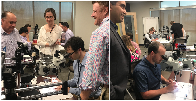
Figure 1 | A camera attached to a small tablet on each microscope provided a video display of what each participant was doing.
A key attribute of this year’s wet lab was the use of a camera attached to a small tablet on each microscope (Figure 1). This enabled a video display of what the participants were doing, facilitating instant feedback and allowing both observers and teachers to view, learn, and teach. In addition to the hands-on experience, the faculty members led discussions over case studies and surgical film review.

The course was concise, covered all available MIGS [devices] with real-life experience, and had enthusiastic support group from Glaukos engineers and researchers.
—Alena Reznik, MD
CONCLUSION
After a stimulating day of lecture and hands-on learning and a facility tour to explore micro-stent manufacturing, we had dinner with the program organizers and cochairs before a final farewell. Attendees were provided with some parting resources so that we could build on our excitement and continue our MIGS education: a copy of Minimally Invasive Glaucoma Surgery: A Practical Guide and a wooden cornice box containing a reusable iPrism surgical gonioscopic lens and iPrism Clip view stabilizer. The iPrism was especially exciting to receive, as it can be sterilized and used in the OR.
All and all, we both agree that the MIGS residents course was a very valuable experience. Packing the lectures and dry lab into 1 day made for a whirlwind, but it seems to be the best way to allow for travel and personal time in a single weekend—not to mention in an ideal geographic location. We cannot wait to return as fellows!


