Supported by


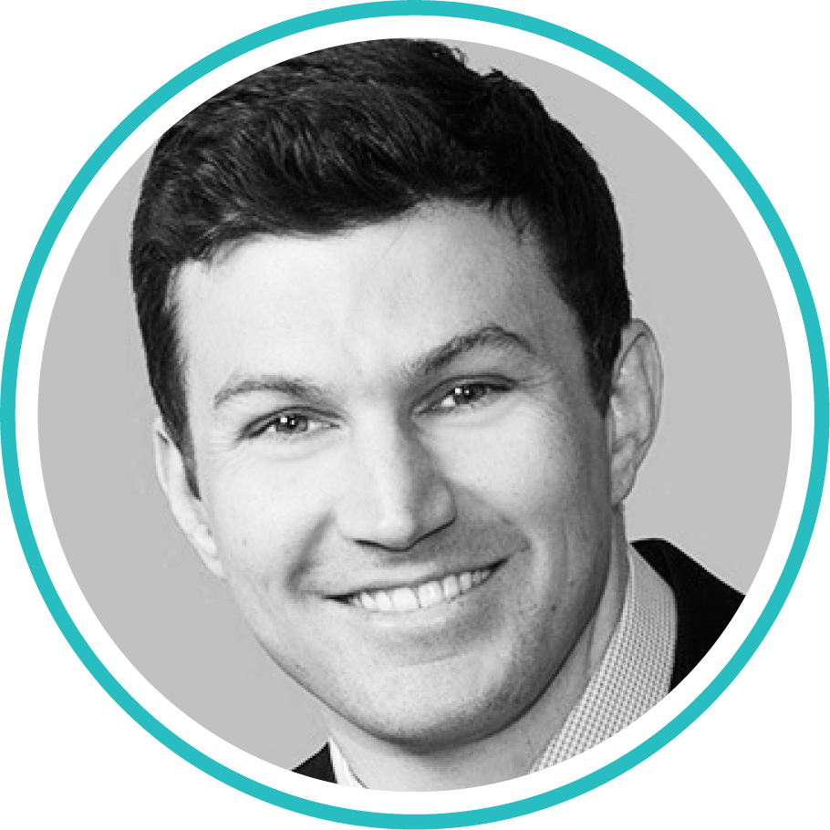
Dr. Duncan is a Cataract, Cornea, and Refractive Surgeon at Horizon Eye Specialists & LASIK Center in Scottsdale, Arizona.
1. Please share with us your background.
I was born and raised in Phoenix, Arizona. While playing football for the University of Arizona, I earned a degree in human physiology with a minor in chemistry. After completing medical school at Midwestern University, I went on to ophthalmology residency at the University of Arizona in Tucson, followed by a fellowship in cornea, refractive, and anterior segment surgery at Baylor College of Medicine in Houston. I currently practice in Scottsdale, Arizona, where I live with my wife and our two daughters.
2. What drew you to ophthalmology and, specifically, to your field of interest?
I was drawn to ophthalmology very early on in my medical career when I realized the impact of restoring someone’s vision. I have continually been amazed by the advancement of technologies in this field and how access to them has translated to better outcomes for my patients. It was the diversity of pathology that drew me specifically to cornea. From complex ocular surface reconstruction, to new and innovative techniques in endothelial keratopathy, to laser vision correction, corneal and refractive surgery offers new and exciting challenges daily.
3. Please describe your current position.
I am a cataract, cornea, and refractive surgeon with Horizon Eye Specialists, a private practice in Scottsdale, Arizona. I have the privilege of working with three other surgeons and six optometrists. We see a very diverse patient population with a broad range of cornea, anterior segment, and refractive pathology at our four offices.
4. Who are your mentors?
Over the years, I have been fortunate to have had many influential mentors. Early on in my career, I was mentored by David Dulaney, MD, who was a pioneer in the field of cataract and refractive surgery. During residency, I learned from Michael Belin, MD, and Mingwu Wang, MD, PhD. Most recently, in my cornea fellowship, I had the opportunity to learn from Doug Koch, MD; Stephen Pflugfelder, MD; Marshall Bowes Hamill, MD; Mitchell Weikert, MD; Sumitra Khandelwal, MD; and Zaina Al-Mohtaseb, MD. Each one is at the top of the field in his or her area of expertise, and the volume and diversity of pathology I was exposed to in Houston made it the perfect environment for training.
5. What has been the most memorable experience of your career thus far?
One of the most memorable experiences of my career was a cataract surgery mission trip to Daet Hospital in the Philippines. I was able to experience what an incredible impact can be made in the lives of patients who have limited access to care. Although we operated on 161 patients over the course of 2 weeks, one patient in particular left a lasting impression. Anna Jane was a very young girl with congenital cataracts. Had it not been for our surgical team, Anna likely would have remained blind for her entire life. While she undoubtedly has some degree of amblyopia, I was touched by the joy and gratitude of her family.
6. What are some new technological advances that you have found particularly exciting? Which advances in the pipeline are you most enthusiastic or curious about?
The advances in presbyopia-correcting IOLs and IOL formulas have been very exciting. Working closely with Dr. Koch, and his vast knowledge in this area, allowed me to fully grasp the complexity of lens calculations and understand how we can potentially improve on this area in the future. I am also excited about the ways corneal crosslinking is evolving, and we are finding new methods of treating corneal ectatic disease—even incorporating it with refractive surgery to achieve better and safer outcomes. I also think endothelial keratoplasty, specifically preloaded Descemet membrane endothelial keratoplasty (DMEK), has been exciting over the past 2 years, and I am enthusiastic about and interested in what the future holds as it relates to cultured endothelial cells for endothelial disease.
7. What is the focus of some of your research?
I have been involved with research in a few different areas, including endothelial keratoplasty and novel secondary IOL fixation techniques. I also worked closely with Dr. Belin on describing the ABCD tomographic staging and classification system for corneal ectasia. We have shown this system, available on the Pentacam (Oculus Optikgeräte), to be a valuable tool in following progression of ectatic disease and monitoring stability after crosslinking.
8. What is a typical day in your life? What keeps you busy, fulfilled, and passionate?
A typical day involves a combination of seeing patients in clinic and performing surgery. As part of a busy anterior segment practice, about 80% of my surgical volume is cataract and refractive surgery, and 20% is cornea and external disease. I typically do two or three DMEK procedures per week, as a significant portion of the corneal disease I see is endothelial dystrophy. Continually striving to help my patients keeps me busy, fulfilled, and passionate. Outside of work, I enjoy spending time with my wife and two girls. I also enjoy biking, hiking, and traveling.
9. What advice can you offer to individuals who are just now choosing their career paths after finishing residency or fellowship?
I would say, above all else, do what makes you happy. For me, I love the diversity of cornea and refractive surgery. I also knew that, when moving to a big city like Phoenix where the job market can be competitive, having additional fellowship training would allow me to have more opportunities. So, for those just finishing training and entering into their careers, decide where you want to live and how to best set yourself apart to be successful there.
10. Tell us about an innovative procedure you are performing or a new imaging/diagnostic tool that has improved your practice.
Several new procedures have really changed my practice. I have transitioned almost exclusively to the Yamane transconjunctival intrascleral fixation technique for secondary IOLs and IOL exchanges without sufficient capsular support. I think this approach offers better and more predictable visual outcomes and shorter recovery than other methods of scleral fixation. Preloaded DMEK has also significantly changed my practice; I have found it to be faster and just as reliable as traditional DMEK.


