


The expanding array of options for surgical correction of presbyopia is certainly to the great benefit of patients, according to Karl G. Stonecipher, MD, clinical associate professor of ophthalmology, University of North Carolina, Chapel Hill, and director of refractive surgery, TLC in Greensboro, N.C.
A device currently in the late stage of US clinical trials (ClinicalTrials.gov Identifier: NCT02374671), the VisAbility Micro-Insert System (Refocus Group) offers a scleral-based approach that appears to restore the patient’s natural accommodative ability. The system consists of four micro-inserts obliquely placed in lamellar, scleral tunnels, 4 mm outside the corneal limbus (Figure). The placement of these micro-inserts restores tension to the zonules, thereby facilitating the ability of the eye to focus on near objects again.
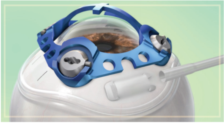
image courtesy of Refocus Group
FIGURE. The placement of these micro-inserts restores tension to the zonules.
“If approved for use in the United States, this system would be the only binocular accommodating restorative procedure that maintains depth of field and depth of focus, features that are not possible with monovision techniques,” Stonecipher says, adding that the system has obtained a CE Mark.
Because the micro-inserts are placed in both eyes, depth of focus and depth of field are maintained. As a result, vision quality at all distances is preserved while near visual acuity is improved. Additionally, the micro-inserts are placed outside the visual axis, thus corneal shape is unaffected and the crystalline lens remains intact. The micro-inserts will not interfere with future refractive procedures and they can be removed if needed.
The procedure is an outpatient surgery typically performed under local anesthesia, and is well within the skillset of any anterior segment surgeon, according to Stonecipher:
- The superior and inferior rectus muscles are marked at the 6-o’clock and 12-o’clock positions, separating the eye into four quadrants
- A 360° peritomy is performed
- A docking station is affixed to the sclera which mates with a scleratome
- Four scleral tunnels are created and the micro-inserts are introduced using a shuttle system
- The conjunctiva is reapproximated; fibrin glue is utilized to maintain hemostasis
This system has generated impressive results in clinical trials in Europe and in studies based in the United States, according to Stonecipher. In a previous multicenter clinical trial of 360 patients, 97% of those who had the bilateral procedure achieved distance corrected near visual acuity at 40 cm of 20/40 (equivalent to J3) or better at 36 months, which was up slightly compared to the 95% noted at 24-months.1 An earlier sample of consecutive cases from two sites in the current US clinical trial (a total of 40 eyes of 20 subjects) visual results showed, 100% of the eyes had uncorrected near visual acuity of 20/40 (J3) or better; 95% were 20/32 (J2) or better and 90% achieved 20/25 (J1) or better with no change to their distance vision at 12 months.
“If it is approved, the system has great potential to revolutionize presbyopia management, offering the potential to restore accommodation in our presbyopic patients while maintaining depth of field and depth of focus without interfering with the visual axis,” Stonecipher says.
1. Katz J. Persistent improvement in near visual acuity 3 years following surgery in the VisAbility multicenter clinical trial. Presented at the American Academy of Ophthalmology. October 15-18, 2016; Chicago.
Dr. Stonecipher reports no relevant financial disclosures.