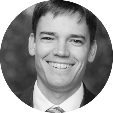
Zachary J. Zavodni, MD
Dr. Zavodni is a cornea, cataract, glaucoma, and refractive surgeon at The Eye Institute of Utah and an Adjunct Clinical Associate Professor at the John A. Moran Eye Center of the University of Utah in Salt Lake City.
Please share with us your background.
I am a first-generation American. My family fled Czechoslovakia upon the Russian takeover after World War II and ultimately landed in Salt Lake City, Utah, where I grew up. As a child, outside of my academic endeavors, my greatest accomplishments were on the tennis court, where I was ranked #1 in Utah for much of my childhood and won a state championship in high school. I attended Northwestern University in Chicago, where I graduated summa cum laude with degrees in biochemistry and economics. Following college, I spent a year conducting macular degeneration research at Duke University in Durham, North Carolina. While there, I met the love of my life, with whom I now have two beautiful children and am celebrating 10 years of marriage this year.
What drew you to ophthalmology and, specifically, to your field of interest?
Like many in medicine, my interest in pursuing a career as a physician grew out of my own experience as a patient. While playing competitive soccer in high school, I was struck in the head by a ball just as it left a defender’s shoelaces. The trauma resulted in a hyphema and multiple retinal tears, requiring many trips to the ophthalmologist. Thankfully, my recovery was uncomplicated, and I regained full vision; I subsequently decided I wanted to one day provide sight restoration to others, as my eye doctor had done for me.
Thanks to a lot of hard work and the help of many mentors along the way, I had the privilege to train as an eye surgeon at Duke University. Early in residency, I found myself gravitating toward anterior segment surgery. I found the endless pursuit for perfection in cataract surgical technique to be addicting. Throw in the analytical mathematical models, which appeal to the engineer in me, and the extraordinarily high rate of life-changing outcomes from sight restoration, and I was hooked on cataract surgery. I was likewise drawn to cornea surgery, in which the quality of a surgeon’s craftsmanship is frequently rewarded with improved visual acuity outcomes for the patient.
Please describe your current position.
I am a junior partner at The Eye Institute of Utah, a medium-sized private practice in Salt Lake City. I provide surgical and medical treatment for all anterior segment subspecialties, including refractive, cornea, glaucoma, and cataract surgery. In addition to operating at our own privately owned ambulatory surgery center, I also spend time as an adjunct faculty member at the University of Utah John A. Moran Eye Center. I hold leadership positions in the American Society of Cataract and Refractive Surgery (ASCRS), including the Young Eye Surgeons (YES) clinical committee. On average, I operate 2 days a week, spend 2.5 days in clinic, and dedicate a half-day per week to my research endeavors.
Who are/were your mentors?
Scott Cousins, MD, has had a profound effect on the trajectory of my career. As a medical student at Duke, he guided me through the design, implementation, and publication of my own clinical research project, which was recognized with a Research to Prevent Blindness (RPB) fellowship award. Dr. Cousins also taught me to recognize the value of my patients as a “living laboratory”—always capable of spurring new clinical questions.
Surgically, I am indebted to Terry Kim, MD; David Hardten, MD; Sherman Reeves, MD, MPH; and Tom Samuelson, MD, who helped me master the fundamentals of cataract, refractive, cornea, and glaucoma surgery. During my anterior segment fellowship at Minnesota Eye Consultants, I was also fortunate to learn the nuances of surgical device innovation from the illustrious Dick Lindstrom, MD (who also happens to be an outstanding tennis player). Finally, I am grateful that my Senior Partner, Robert Cionni, MD, continues to mentor me every day, both as a surgeon and a businessman.
What has been the most memorable experience of your career thus far?
I hate to say it, but my most memorable experiences to date have been my cases that have—for some reason or another—had poor outcomes. Thankfully, there have not been many, but I have certainly had a few sleepless nights following a tough day in the OR. While unpleasant, these growing pains have unquestionably made me a better surgeon. I make a point to review my surgical cases with my senior partners, and doing so has equipped me with an ever-growing toolbox of surgical techniques as well as a subdued self-confidence that I believe benefits my patients.
What are some new technological advances that you have found particularly exciting? Which advances in the pipeline are you most enthusiastic or curious about?
As a frequent user of femtosecond laser cataract surgery, I recognize many of its advantages and disadvantages. While the reproducibility and uniformity of laser capsulotomy creation is a luxury, the frequent pupil dilation difficulties and sticky cortex adhesions at the capsulotomy edge are not ideal. Consequently, I am excited about the early results of the new Zepto system (Mynosys), which employs a novel technology coined precision pulse capsulotomy.
The Zepto is a disposable, foldable device that can fit through a 2.2-mm incision and, upon suction to the anterior capsule, can create a perfect capsulotomy using a series of microsecond-long electrical pulses. The beauty of Zepto is that it can be integrated as an additional step during routine phacoemulsification, it can be centered on the Purkinje image, it can easily be employed following the insertion of an iris-retracting device, it has not been associated with resultant miosis, it does not create sticky cortical attachments at the rhexis edge, and it is significantly cheaper than conventional laser cataract surgery. The upcoming clinical trials should be very interesting to watch unfold.
What is the focus of some of your research?
The lion’s share of my research these days is focused on corneal collagen crosslinking (CXL) and its ability to arrest ectatic processes. I have been involved in multiple protocols, including Crosslinking USA, Avedro’s phase 3 randomized controlled trial, and, most recently, our practice’s own protocol directly comparing epithelium-on with epithelium-off techniques. I was thrilled by Avedro’s announcement of FDA approval in April, as it is the first step toward easier access to this essential technology. With approval, I am also hopeful that US surgeons will begin to explore the efficacy of CXL combined procedures, such as CXL with intrastromal corneal ring segments (ie, Intacs; Addition Technology) and CXL with PRK (sadly, we are far behind our European and Canadian counterparts in this regard).
What is a typical day in your life? What keeps you busy, fulfilled, and passionate?
I feel fortunate that I get excited driving to work. Whether it’s a full day of clinic or in the OR, I love the challenges, successes, and questions that each day brings. While my clinical duties encompass the majority of my time, I find research to be an invaluable part of my job, as research lays the foundation for true innovation and change. I am also passionate about teaching. We have many medical students and residents rotate through our clinic, and watching them develop the skills to make both surgical and management decisions on their own is tremendously rewarding.
When I am not at work, I am at home with my wife and our two beautiful children, ages 6 and 2. There is nothing more humbling and rewarding than being a parent. There is also nothing that motivates me to succeed in my profession more than their smiling faces, sending me off every morning and greeting me each evening.
What advice can you offer to individuals who are just now choosing their career paths after finishing residency or fellowship?
The first few years out of residency and fellowship can be tough, especially in the OR as you adjust to not having an attending present to back you up. Inevitably, some cases are not going to go the way you had hoped or watched on the big screen at national meetings. The key to maintaining your confidence and improving surgically during this integral time is to recognize you are not alone. Don’t ever hesitate to reach out to your old residency or fellowship mentors for advice about a tough case. Not only is it an opportunity to learn, but also actually a great way to maintain friendships.
I would also strongly recommend when you are looking for your first job to try and find at least one, if not more, senior partner who genuinely wants to work closely with you and teach you everything he or she knows. Make a point to review surgical videos together on a regular basis. If you can find these assets in your first job setting, you will quickly gain confidence and your surgical skillset will expand at an impressive rate.
Tell us about an innovative procedure you are performing or a new imaging/diagnostic tool that has improved your practice.
While not especially new, wavefront intraoperative aberrometry has been an invaluable tool in our practice for the past several years. When I go to national meetings, I am consistently surprised by the lack of adoption of this technology. It has been particularly useful in helping to nail down the IOL choice for our post-refractive surgery patients. In our hands, the ORA System (Alcon) helps us deliver a final refraction within 0.50 D of target more than 80% of the time in post-myopic LASIK eyes.
In addition to intraoperative aberrometry, I would be remiss not to mention a new and elegant sutureless technique for IOL scleral fixation that I came across at this years’ ASCRS meeting. The technique employs the use of a three-piece IOL with externalization of the haptics through transconjunctival 30-gauge needle tracks in the posterior sulcus plane. The haptics are secured by simply melting the tip of the haptic with a handheld cautery and creating a bulb larger than the needle track. The ends are tucked into the scleral tunnels, and no conjunctival incisions or glue are required. The technique was introduced by Shin Yamane, MD, of Yokohama, Japan, and won the Grand Prize at the ASCRS Film Festival Awards. To date, we have attempted this technique on approximately10 patients in the practice and had outstanding results. I would encourage everyone to review this video.


