


"This patient also has pseudoexfoliation," described Matt Oliva, MD, as I peered over his shoulder to observe one of the 271 cataract operations that he performed over 3.5 days during a Himalayan Cataract Project (HCP) surgical outreach campaign in Hosanna, Ethiopia. As a second-year ophthalmology resident, I was thrilled to be invited to learn about outreach camp logistics, surgical techniques, and a patient population I do not commonly encounter at my home institution.
As Dr. Oliva continued to point out patient after patient with pseudoexfoliation syndrome (PXF) on the operating table, I became intrigued by the high prevalence of PXF in the Ethiopian population. Classically, residents are taught that PXF is most commonly associated with Scandinavian heritage. However, the prevalence of PXF in Ethiopian patients with cataracts was reported to be 39%1 and 36%2 in two studies, whereas a recent population-based evaluation in central Ethiopia found a prevalence of 13% in patients who were 40 years of age or older.3 The prevalence of PXF varies widely, but these proportions are staggering compared to the rates reported in South Korea for patients who are 50 years of age or older (0.12%)4 and in the Southeastern United States for patients who are 30 years of age or older (1.6%).5 The incidence of PXF in Ethiopians is, in fact, comparable to that reported in people of Scandinavian descent: 11% in Iceland,6 22% in Finland,7 and 23% in Northern Sweden.8
Within Africa, the prevalence of PXF varies considerably, but Ethiopia has one of the highest rates of PXF, as summarized in a systematic review conducted by Olawoye et al in 2014.9 Studies of PXF in South Africa found a prevalence that ranged from 5% to 16% depending on ethnic group, and clinic-based studies in Ghana, Nigeria, and the Democratic Republic of the Congo reported a prevalence of PXF of 0%, 3%, and 2%, respectively.9 The high prevalence of PXF in people with cataracts may reflect the propensity of individuals with PXF to develop cataracts, as described in the literature, and may partly explain the high cataract burden in Ethiopia.
PXF AND CATARACT SURGERY
When it comes to cataract surgery, PXF is known to increase the risk of complications by causing pupillary miosis and zonular instability.10 Surgeons prepare for the worst when planning phacoemulsification on patients with PXF, and they have devised a variety of tips and tricks to use in high-resource settings.10,11
In Hosanna and other low-resource settings, however, cataracts are often removed with manual small-incision cataract surgery (MSICS) instead of phacoemulsification. MSICS is a modified extracapsular technique that involves creating a self-sealing, 7- to 8-mm sclerocorneal tunnel incision through which the nucleus is delivered whole. The instruments required for this approach are inexpensive.
A Cochrane review reported similar visual outcomes and complication rates with MSICS and phacoemulsification, although the studies included were underpowered and had short follow-up periods.12 Other attempts to compare MSICS and phacoemulsification in eyes with PXF have been similarly limited by small sample sizes and short follow-up periods.13,14
PXF AND MSICS 101
PXF challenges both MSICS and phaco surgeons. As a budding cataract surgeon with little exposure to MSICS, I was curious about how to approach the procedure in patients with PXF. I learned a lot about managing small pupils, phacodonesis, and lens subluxation or dislocation from David Khorram, MD, and Allison Jarstad, DO. Dr. Khorram is the director of global ophthalmology at the University of Virginia and works regularly with the HCP in Ethiopia. Dr. Jarstad is a cornea and refractive surgery specialist in Southern California and the 2018 to 2019 HCP/Stanford University Global Ophthalmology Fellow.
Pupil Expansion
Adequate pupil expansion is important when performing MSICS because a small pupil can make it difficult to prolapse the nucleus into the anterior chamber, potentially increasing stress on already weakened zonules. In a high-resource setting, the pupil can be expanded with viscodilation, mechanical stretching, iris hooks, or devices such as the Malyugin Ring (MicroSurgical Technology).
In Hosanna and other low-resource settings, iris sphincterotomies are the primary means of expanding the pupil. For this approach, Dr. Jarstad recommends that the surgeon “insert long Vannas scissors through the main wound incision and create eight 1-mm sphincterotomies that are evenly spaced.” Although this may raise concern about disfigurement, the pupil tends to return to a smaller size postoperatively. Additionally, posterior synechiae are more likely to be present in eyes with PXF, and synechiolysis may be required. This can be performed with an OVD, a blunt instrument, or long Vannas scissors.
Capsulotomy Creation
Zonulopathy may result in phacodonesis, which requires the MSICS surgeon to adopt a gentle approach. As the nucleus must be prolapsed during MSICS, a generous 7.0-mm anterior capsulotomy is recommended. Because it is challenging to construct a large continuous curvilinear capsulorhexis (CCC), Dr. Jarstad prefers to create a V-shaped or smile-shaped anterior capsulotomy. This approach helps to ensure that the IOL will be inserted into the bag because the capsular flap—folded anteriorly over the iris—will block the leading haptic of an IOL from being inadvertently introduced in the sulcus. Similarly, Dr. Khorram favors an envelope capsulotomy. With all of these capsulotomy techniques, it is important to excise the flap after IOL insertion.
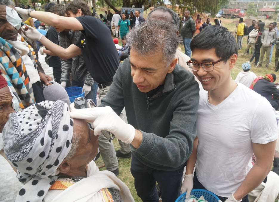
Figure. Drs. Lau and Khorram during a high-volume cataract campaign. Image courtesy of Christopher Briscoe.
Other advantages of performing one of these capsulotomies instead of a CCC are that the former can be constructed when there is no red reflex and the capsulotomy can easily be enlarged to accommodate a large nucleus. In addition, these capsulotomies are quick to create, and their ability to expand as needed reduce the stress placed on the zonules when prolapsing a bulky nucleus. In a low-resource setting, the surgeon’s choice of capsulotomy technique may be influenced by the instruments available in the field.
Nucleus Prolapse and Delivery
When prolapsing the nucleus, Dr. Jarstad recommends performing gentle hydration with a Simcoe cannula pointed posteriorly and toward one side. This maneuver causes the opposite pole of the nucleus to prolapse anteriorly, and, afterward, the surgeon can gently sweep the Simcoe cannula under the center of the nucleus to prolapse the rest of the nucleus into the anterior chamber. The nucleus may then be delivered with the Simcoe cannula, a Vectis lens loop, or the fishhook technique.
In the fishhook technique, after the superior pole of the nucleus has prolapsed into the anterior chamber, an OVD is injected posterior to the nucleus to separate it from the posterior capsule and underlying iris. A 26- or 27-gauge needle with its tip bent in the shape of a fishhook is inserted via the main incision such that the fishhook tip is parallel to the iris plane. The needle is slid to the center of the nucleus, at which point the needle is rotated 90 so that the tip faces anteriorly and impales the nucleus. The nucleus is then delivered by pulling the needle with the nucleus out of the main wound. In Dr. Jarstad’s experience, especially when the nucleus is bulky, extracting the nucleus is easier with this technique than with the larger Vectis lens loop or a Simcoe cannula, owing to the small profile of the fishhook needle.
Dr. Khorram follows the same principles to ensure prolapse and removal of the nucleus with minimal stress on the zonules, but he uses a different technique. “Prolapsing the nucleus can be one of the most challenging steps of learning MSICS,” explained Dr. Khorram. “I place the Simcoe cannula wide open beneath the anterior capsular flap to perform hydrodissection. I then remove the cannula; infraduct the eye; and with the Simcoe on the external lip of the incision and slight posterior pressure, pop the superior nucleus into the anterior chamber through a generous capsulotomy.” Dr. Khorram then uses the irrigation of the Simcoe cannula to bring the nucleus into the anterior chamber, places the cannula beneath the nucleus, and pulls it out of the eye with a combination of the hydrostatic and mechanical forces of the cannula. “It is a gentle and reliable technique that I learned from Dr. Oliva,” said Dr. Khorram.
IOL Insertion
After nuclear extraction and cortical cleanup, Dr. Jarstad typically inserts a three-piece IOL in the bag or in the sulcus if reasonable capsular support is present. If a CCC has been created, ideally, optic capture of the IOL will be performed. If there is no capsular support, she recommends implanting an anterior chamber IOL (ACIOL), in which case a peripheral iridotomy is required to prevent pupillary block.
Lens Subluxation
In an eye with severe zonulopathy, the cataractous lens may subluxate, in which case the principles described earlier still apply. Depending on the amount of zonular laxity, one may attempt either an intracapsular or extracapsular technique. For cases in which the lens has not subluxated severely and no vitreous is present in the anterior chamber, Dr. Khorram performs an envelope capsulotomy and removes the nucleus with a Simcoe cannula.
Dr. Jarstad describes an alternative approach. A CCC is preferable to another capsulotomy technique in this situation due to the higher risk of radialization when creating a capsulotomy at the lens periphery. Dr. Jarstad recommends using an OVD after hydrodissection to deliver the nucleus slowly and gently into the anterior chamber. If too much zonular dialysis is observed after nuclear removal to permit safe cortical cleanup or the implantation of a posterior chamber IOL, Dr. Khorram will remove the capsular bag with all of the cortical remnants.
However, for most cases of significant subluxation, Dr. Khorram recommends an intracapsular technique to prevent the nucleus from dropping into the vitreous, which may result in vigorous lens-induced posterior segment inflammation and retinal damage. This is of particular concern in low-resource settings where vitreoretinal surgery support is not available. When using an intracapsular technique, Dr. Khorram recommends creating a similar sclerocorneal tunnel incision, injecting adequate OVD posterior to the capsular bag for support, and moving the entire lens into the anterior chamber, either with an OVD cannula or with a Vectis lens loop. The tunnel incision should be enlarged to accommodate safe removal of the entire lens from the eye.
CONCLUSION
Surgeons who volunteer their time and skill to treat patients in low-resource settings such as Ethiopia deserve the utmost respect. I was stunned to see the surgical complexity they face regularly in this pursuit. Although small pupils and zonulopathy are common in patients with PXF, these challenges can be encountered in any cataract case. Tackling these problems with limited resources is particularly difficult, but the principles outlined in this article should serve those who are combatting cataract blindness in all settings.
1. Teshome T, Regassa K. Prevalence of pseudoexfoliation syndrome in Ethiopian patients scheduled for cataract surgery. Acta Ophthalmol Scand. 2004;82:254-258.
2. Gelaw Y, Tibebu Y. Clinical characteristics of cataract patients with pseudoexfoliation syndrome at Jimma University Specialized Hospital, Southwest Ethiopia. Ethiop J Health Sci. 2012;22(1):1-6.
3. Berhanu Y, Giorgis A, Alemu A. Prevalence of ocular pseudoexfoliation in Baso and Worena District, central Ethiopia. Ethiop J Health Dev. 2020;34(1):54-58.
4. Soa K, Su-Ho L, Kyung RS, et al. Prevalence of pseudoexfoliation syndrome and associated factors in South Koreans: The Korean National Health and Nutrition Examination Survey. Ophthalmic Epidemiology. 2016;23(5):298-302.
5. Cashwell LF Jr, Shields MB. Exfoliation syndrome in the Southeastern United States I. Prevalence in open-angle glaucoma and non-glaucoma populations. Acta Ophthalmol. 1988;66(S184):99-102.
6. Arnarsson AM. Epidemiology of exfoliation syndrome in the Reykjavik Eye Study. Acta Ophthalmol. 2009;87(3):1-17.
7. Hirvela H, Luukinen H, Laatikainen L. Prevalence and risk factors of lens opacities in the elderly in Finland. A population-based study. Ophthalmology. 1995;102(1):108-117.
8. Astrom S, Linden C. Incidence and prevalence of pseudoexfoliation and open-angle glaucoma in northern Sweden: I. baseline report. Acta Ophthalmol Scand. 2007;85(8):828-831.
9. Olawoye OO, Pasquale LR, Ritch R. Exfoliation syndrome in sub-Saharan Africa. Int Ophthalmol. 2014;34(5):1165-1173.
10. Calafati J, Tam DY, Ahmed IK. Pseudoexfoliation syndrome in cataract surgery. Eyenet Magazine. April 2009. www.aao.org/eyenet/article/pseudoexfoliation-syndrome-in-cataract-surgery. Accessed July 4, 2020.
11. Nazarali S, Damji F, Damji KF. What have we learned about exfoliation syndrome since its discovery by John Lindberg 100 years ago? Br J Ophthalmol. 2018;102:1342-1350.
12. Riaz Y, de Silva SR, Evans JR. Manual small incision cataract surgery (MSICS) with posterior chamber intraocular lens versus phacoemulsification with posterior chamber intraocular lens for age-related cataract. Cochrane Database Syst Rev. 2013;10:CD008813.
13. Naik AU, Gadewar SB. Visual outcome of phacoemulsification versus small incision cataract surgery in pseudoexfoliation syndrome – a pilot study. J Clin Diagn Res. 2017;11(1):NC05-NC08.
14. Dash N, Choudhury D. Is manual small-incision cataract surgery (MSICS) a better option than phacoemulsification for managing pseudoexfoliation cataracts in a developing nation? Poster presented at: ESCRS Annual Meeting; September 14-18, 2019; Paris, France.


