


Glaucoma is the leading cause of irreversible blindness worldwide.1 It is therefore important to establish cost-effective and accurate methods of screening patients for the disease in settings with limited resources. Two studies of the prevalence of glaucoma in Nepal suggested that screening people for the disease using a combination of portable instruments and teleophthalmology may be an effective strategy for identifying patients with glaucoma in remote locations.
The Bhaktapur Glaucoma Study (BGS)2 and Jiri Eye Study (JES; S.E. Miller, N. Blackburn, S.S. Thapa, et al, unpublished data, 2021) were conducted in different geographic locations of Nepal. These population-based studies estimated an overall prevalence of glaucoma of 1.6 % and 4.2%, respectively. In both studies, the prevalence of primary open-angle glaucoma (POAG) was greater than that of primary angle-closure glaucoma (PACG):
- POAG 1.2% and PACG 0.4% in the BGS; and
- POAG 4.2% and PACG 0.8% in the JES.
More than 90% of the individuals with glaucoma were unaware of the disease and diagnosed for the first time in these studies. Glaucoma was the cause of blindness in 5% and 0.2% of the blind individuals in the BGS and JES, respectively.
Although population-based programs are not a cost-effective way to screen for glaucoma,3 findings from the BGS and JES suggested ocular parameters that can be used for disease screening. In both studies, more than 70% of individuals with glaucoma had normal IOP, but more than 75% exhibited changes in the optic disc and visual field (VF) at the time of diagnosis. These findings indicate that screening people for glaucoma by examining the optic disc and VF rather than by measuring IOP could be an effective approach.
CONSIDERATIONS IN NEPAL
The clinic is the preferred setting for detecting glaucoma.3 Although it is important to diagnose glaucoma (especially moderate to advanced disease that could cause blindness during the patient’s lifetime), it is also important to avoid referrals of erroneously identified glaucoma suspects. The latter is of particular concern in developing countries such as Nepal where people must travel for several days across harsh terrain to reach an eye care facility. A lack of trained personnel and slowly developing infrastructure for eye health systems are additional challenges.4,5
To overcome these barriers, ophthalmic technicians have begun traveling to remote locations, using inexpensive portable instruments (such as fundus cameras and VF analyzers), and implementing teleophthalmology consultations with an ophthalmologist. A recent study evaluated the ability of a suprathreshold perimetry app on an iPad (Apple) to detect moderate to advanced glaucoma.6 The software detected most moderate (mean deviation [MD], -6 to -12 dB) and advanced (MD, > -12 dB) VF deficits. The app was less accurate at detecting early (MD, < -6 dB) disease, primarily because of an elevated false-positive response rate.
The use of virtual perimetry for identifying glaucomatous VF defects is also being studied (Figure 1). After adjusting for the grader and measurement order, no significant difference was found in cup-to-disc measurements obtained using images taken with a portable nonmydriatic fundus camera versus a traditional tabletop mydriatic fundus camera.7 Two studies subsequently conducted at community eye centers and screening camps in Nepal (Figure 2) compared screening referral recommendations made by remotely located ophthalmic technicians with those of an ophthalmologist reviewing digital photographs obtained with a portable ophthalmic camera system powered by a mobile device that were transmitted through a teleophthalmology system.8,9
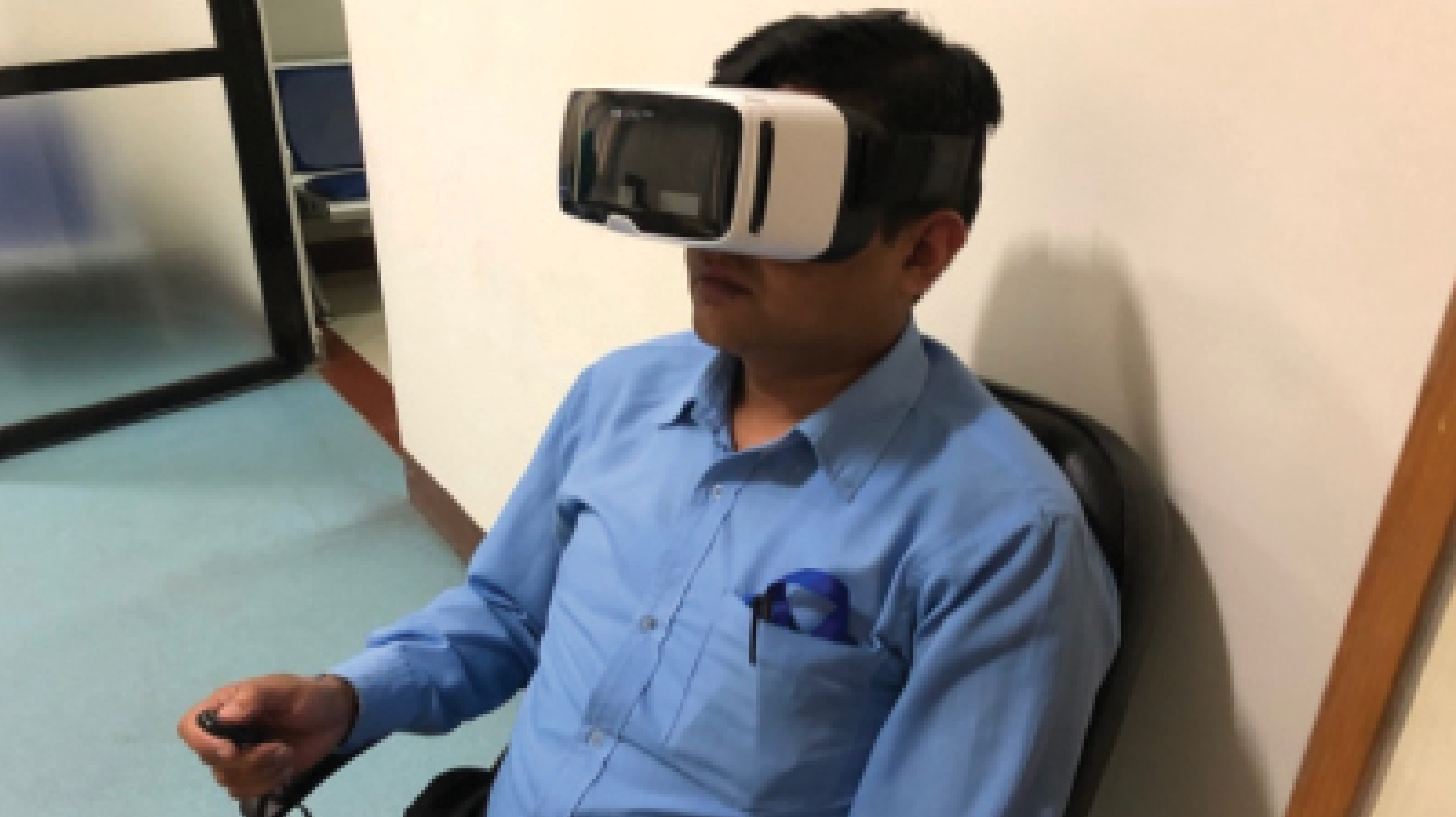
Figure 1. A patient undergoes a visual field examination using virtual reality perimetry.
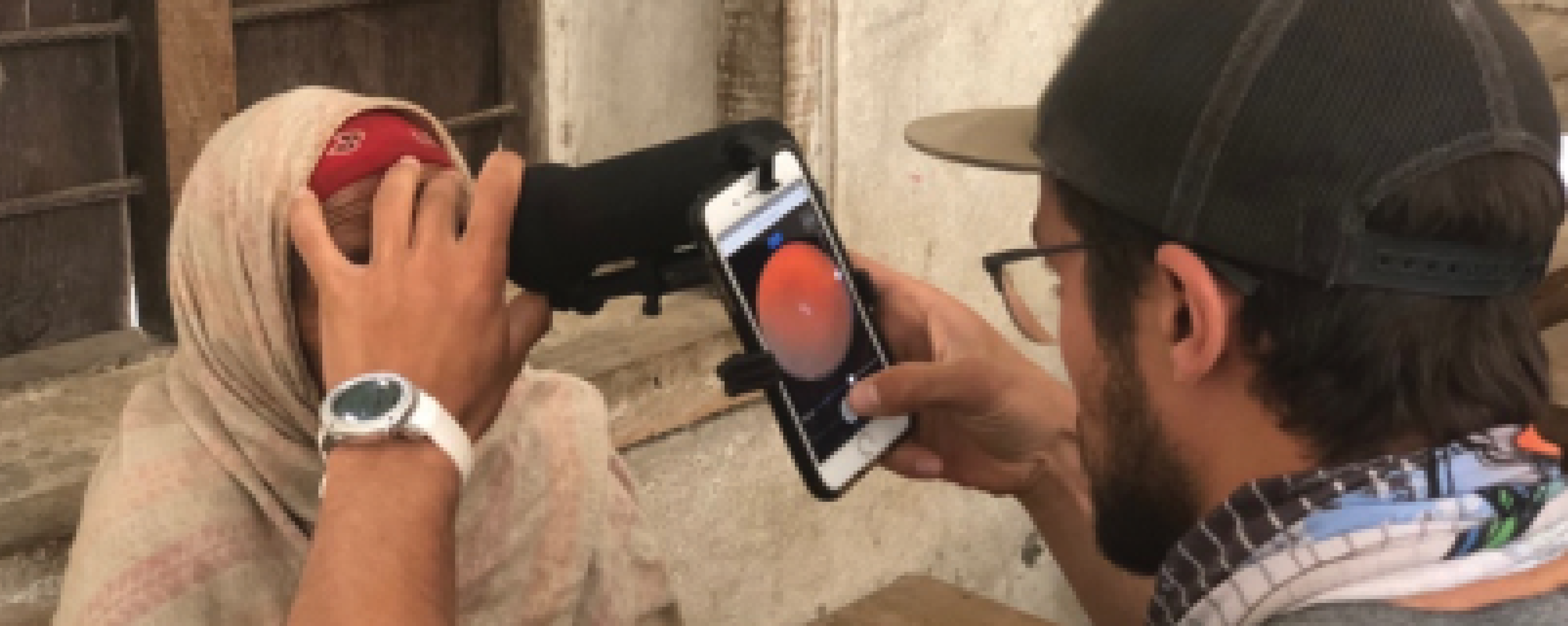
Figure 2. A mobile device and teleophthalmology system are used to screen patients in remote regions of Nepal.
In both studies, the ophthalmologist identified a significant number of patients whose need for a referral had not been identified by the screening technician. These results suggest that teleophthalmology using a mobile device can serve as an important adjunct to patient care being delivered by technicians at community eye centers and screening camps in rural Nepal. Combining technologies to estimate cup-to-disc ratio and detect VF defects with clinical examinations performed by an ophthalmic technician could increase the chance of diagnosing glaucoma accurately.
CURRENT EFFORTS BY THE TILGANGA INSTITUTE OF OPHTHALMOLOGY
The Tilganga Institute of Ophthalmology has 25 community eye centers in various remote locations of Nepal. They are run by ophthalmic technicians. Recently, 10 ophthalmic technicians working at these centers were provided with a mobile device and teleophthalmology system. They have begun obtaining and transmitting fundus photographs to an ophthalmologist located in Kathmandu.
The criteria for a teleophthalmology consultation are as follows:
- Visual acuity of less than 6/12 with no media opacity that does not improve with pinhole;
- Retinal pathology; and
- Suspicious or glaucomatous optic disc.
Challenges encountered thus far include the COVID-19 pandemic, monitoring the efficiency with which ophthalmic technicians use mobile devices and the teleophthalmology system, and keeping these employees motivated. Time is another limiting factor because the remote screening clinics are attended by a large number of patients.
It is to be hoped that accurate, cost-effective systems for glaucoma diagnosis and patient referral can be established in remote locations of Nepal in the near future.
1. Bourne RRA, Stevens GA, White RA, et al; Vision Loss Expert Group. Causes of vision loss worldwide, 1990–2010: a systematic analysis. Lancet Glob Health. 2013;1(6):e339-e349.
2. Thapa SS, Paudyal I, Khanal S, et al. A population-based survey of the prevalence and types of glaucoma in Nepal: the Bhaktapur Glaucoma Study. Ophthalmology 2012;119(4):759-764.
3. Thomas R, Paul P, Muliyil J. Glaucoma in India. J Glaucoma. 2003;12(1):81-87.
4. Thomas R, Kumar RS, Chandrasekhar G, Parikh R. Applying the recent clinical trials on primary open angle glaucoma: the developing world perspective. J Glaucoma. 2005;14(4):324-327.
5. Thylefors B. A global initiative for the elimination of avoidable blindness. Indian J Ophthalmol. 1998;46(3):129-130.
6. Johnson CA, Thapa S, George Kong YX, Robin AL. Performance of an iPad application to detect moderate and advanced visual field loss in Nepal. Am J Ophthalmol. 2017;182:147-154.
7. Miller SE, Thapa S, Robin AL, et al. Glaucoma screening in Nepal: cup-to-disc estimate with standard mydriatic fundus camera compared to portable nonmydriatic camera. Am J Ophthalmol. 2017;182:99-106.
8. Hong K, Collon S, Chang D, et al. Teleophthalmology through handheld mobile devices: a pilot study in rural Nepal. J Mob Technol Med. 2019;8(1):10.7309/jmtm.8.1.1.
9. Collon S, Chang D, Tabin G, et al. Utility and feasibility of teleophthalmology using a smartphone-based ophthalmic camera in screening camps in Nepal. Asia Pac J Ophthalmol (Phila). 2020;9(1):54-58.
