
Stuart Stoll, MD
SimulEYE Ophthalmic Surgical Training Devices
After completing a LASIK-only fellowship and not performing cataract surgery for a year, I found that the learning curve back into the eye was steep. Therefore, I sought a way to practice the most difficult step of cataract surgery, the continuous curvilinear capsulorhexis (CCC), to quickly flatten the learning curve. The available options for practicing the CCC technique were grapes, messy and unrealistic pig eyes, and a couple of inadequate artificial eyes. A colleague of mine recommended the use of cellophane wrapping paper, which, while lacking in realism, was nonetheless effective for practicing CCC tears and understanding the directional forces at work. Following that experience, the SimuloRhexis CCC training device was subsequently born as a rudimentary handheld device using Silly Putty and cellophane. It has since undergone many generational advances to evolve into the robust and realistic device that it is now. From there, I recognized the need to provide realistic training devices for other aspects of ophthalmic surgery, leading to the development of the SimulEYE line of ophthalmic surgical training devices.
Recognizing that patients present with very different ocular conditions and anatomy, it is not possible to create one artificial training eye that allows surgical training across a variety of conditions. Each device in the SimulEYE line is designed to target a specific surgical procedure and homes in on that technique with as much realism as possible. The external housing was designed to allow multiple components to be placed inside to allow training in a variety of situations. In addition, the suction cup base allows the devices to be used on a flat surface, such as on a desk for viewing through a surgical microscope or on a vertical stand to practice other laser procedures. A flexible stalk just above the suction cup base allows for realistic eye movements during surgical training. The footprint is extremely small and does not require any additional supporting equipment other than a slit-lamp stand for certain laser procedures. The SimulEYE devices are single-use or multi-use, depending on the version and the technique being demonstrated.
SimuloRhexis
SimulEYE SimuloRhexis, more easily referred to as SimuloRhexis, allows the user to practice up to seven different capsulotomies per eye while providing a progressively more realistic and challenging environment. The capsule of SimuloRhexis has extraordinarily realistic tearing properties and a feel that closely mimics the human anterior capsule. The surgeon begins with a revolver template in place, which acts as a guide to create six peripheral CCCs. These are created with the use of viscoelastic placed over each opening immediately prior to initiating the tear. The viscoelastic must be placed generously to create a dome, which acts as the anterior chamber and allows the capsule to float within it for realistic manipulation. The surgeon can work within one of three gates, which act as an incision guide while not restricting movement as much as an actual incision. The other three tears can be performed over the bars that join the gates, and this creates a different angle of approach to the CCC, as might be encountered in a deeper chamber. Upon completing the six peripheral tears, the revolver template is removed and replaced by the central pupil and the cornea cap. The central film has been preserved by the revolver template and is now available for one final attempt under the most realistic conditions. Incisions are made in the cornea, the anterior chamber is filled with viscoelastic, and a central capsulorhexis is performed. For greater realism and difficulty, the posterior pressure can be dialed up, which will cause the chamber to shallow and the anterior capsule to bow upward. The CCC tear will have a tendency to run outward, and the rescue technique can be practiced.
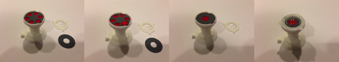
SimuloRhexis components and assembly for use.
A video demonstration of SimuloRhexis features and use.
SIMULEYE YAG, SIMULEYE SLT
SimulEYE YAG and SimulEYE SLT allow for training on YAG and selective laser trabeculoplasty (SLT) lasers so that the user can become intimately familiar with these devices in terms of settings, features, and surgical technique prior to working with an actual patient. SimulEYE YAG provides an aqueous environment with an anterior chamber, iris, anterior capsule overlying an IOL, and a posterior capsule. The YAG laser is used to focus through the IOL and open the posterior capsule. Focusing, aiming beam, and power adjustments along with offsets can be changed to see the effects inside the eye so that the user can learn the instrument and avoid pits in the IOL while creating an effective opening in the posterior capsule.
SimulEYE YAG video demonstration. The pupil margin can be visualized as well as the margin of the IOL and an opacified posterior capsule. An Nd:YAG laser is used to focus through the IOL and deliver energy to the posterior capsule while avoiding pitting of the IOL.
In a similar way, SimulEYE SLT allows for training with the SLT laser. This model provides an aqueous environment and a pigmented trabecular meshwork, which acts as the laser target. The use of an SLT laser lens with a coupling agent is required and helps the user to understand the principles of treating the angle. When targeting the pigmented trabecular meshwork, power settings can be adjusted to demonstrate formation of champagne-like bubbles at higher energy levels, which disappear at lower power settings. A robust pigment change is designed to occur with each pulse so that the user can identify the treated area. This helps the surgeon learn how to properly rotate the mirrored lens, locate the area that has been treated, and continue the treatment in the desired area.
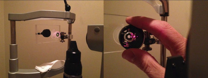
SimulEYE SLT vertically mounted on a slit-lamp stand so that the eye is in the proper position for treatment. An SLT laser lens is applied for viewing and treating the angle.
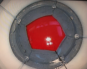
SimulEYE SLT video showing a gonio mirror view of the pigmented trabecular meshwork laser target. SLT laser delivery causes a depigmentation for easy visualization of treated areas. Champagne-like bubbles can be visualized with each laser pulse and will increase or decrease accordingly as energy levels on the laser are adjusted.
SimulEYE SMALL PUPIL
SimulEYE Small Pupil has been designed with a flexible iris to help train surgeons in the use of various pupil expansion techniques. Surgeons can practice by using two instruments in a push-pull technique, placing iris hooks, or using one of several iris expander devices.
SimulEYE Small Pupil being used with iris hooks. Four MST iris hooks and one Alcon/Grieshaber iris hook have been placed to open the pupil.
SimulEYE Small Pupil video demonstrating the use of the MST Malyugin Ring. Various techniques of implantation and removal can be practiced, and the eight points of fixation are demonstrated when the ring is in place.
SimulEYE Aphakia used in conjunction with the Zeiss Callisto system. The model provides an empty capsular bag with a premade 5.5-mm capsulorhexis for implantation and rotation of a toric IOL.
Tracking of the SimulEYE model. The yellow registration line and the blue toric IOL location line remain stable during eye movement.
MODELS IN DEVELOPMENT
Several other models in the SimulEYE line are currently in development.
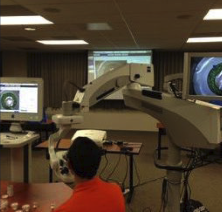
• SimulEYE Femto allows demonstration of femtosecond lasers for cataract surgery. Features of the femtosecond laser platforms that can be demonstrated include docking, registration, auto focus, optical coherence tomography imaging, and identification of anatomic landmarks along with a realistic treatment effect when firing the femtosecond laser.
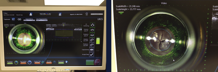
SimulEYE Femto on the Victus laser platform (Bausch + Lomb). OCT imaging demonstrates anatomic landmarks, including the cornea, anterior chamber, iris, and the anterior and posterior capsules of the lens (Left). Fragmentation pattern in the lens and the resultant bubbles in the anterior chamber can be seen after treatment with the Victus laser (Right).
• SimulEYE Retinal Laser is similar to the YAG and SLT versions in that it can be mounted using the slit-lamp stand. This model will allow practice with the multispot pattern photocoagulation lasers.
• SimulEYE Anterior Segment will allow more advanced anterior segment surgeons to practice techniques of IOL and iris suturing along with pars plana anterior vitrectomy.
• SimulEYE PPV will provide an environment for the vitreoretinal surgeon to practice vitrectomy techniques and learn membrane peeling.
• SimulEYE Phaco will provide the most realistic and comprehensive approach to practicing phaco cataract surgery.
SimulEYE Femto used to demonstrate the Alcon LenSx femtosecond laser platform. OCT imaging shows the anatomic landmarks. A realistic treatment effect is seen when firing the laser. Capsulotomy, lens fragmentation, and all corneal incisions can be visualized.
The realistic and versatile platform of the SimulEYE line of devices has allowed the models to be used effectively in resident training, exhibition of state-of-the-art technology, and advanced training of experienced surgeons. SimulEYE provides realism and repeatability in a small footprint, which is cost effective, and without the sterility and disposal issues of animal eyes.
TRAIN
SimulEYE models have been used in the CORE resident program along with other multiresidency training programs. The ability of a resident surgeon to be able to practice techniques multiple times prior to live surgery is invaluable, especially when performed under the direction of an experienced surgeon.
EXHIBIT
As the field of ophthalmology is so highly device- and technology-dependent, there has been considerable interest from the ophthalmic industry in the use of SimulEYE to showcase technology and train surgeons on various devices.
ADVANCE
Even established ophthalmic surgeons have asked for and been able to benefit from the SimulEYE line of devices. Advanced intraocular skills such as IOL suturing and various other skills that are not routinely encountered can be learned or refreshed prior to live surgery.
CONCLUSION
The SimulEYE line of ophthalmic training devices, while only recently launched in select markets, is gaining in popularity and acceptance. SimulEYE will continue to advance and improve to meet the growing needs of the ophthalmic community.
Stuart Stoll, MD, is an ophthalmologist at Beverly Hills LASIK Center in Beverly Hills, California. Dr. Stoll is the Founder of insEYEt LLC and Creator of the SimulEYE ophthalmic surgical training devices. He may be reached at stustoll@gmail.com.
For more information or to order the SimulEYE devices, visit.www.SimulEYE.com (currently under construction) or contact info@insEYEt.biz


