supported by


Olivia L. Lee, MD
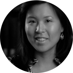
Olivia L. Lee, MD, is an Assistant Professor of Ophthalmology at the Doheny Eye Institute, David Geffen School of Medicine at UCLA, in Pasadena, California.
1. Please share with us your background.
I was born and raised in Maryland, the first-born child of immigrants. My parents came to the United States to pursue graduate school and decided to stay so that I could grow up here. From the day I was born, my parents told me I would go to law school one day. As a child, I loved crafts, and my grandmother taught me to paint, but my parents instructed me to save these hobbies for retirement. With filial piety engrained in me, I obeyed my parents until a brief experience on the mock trial team in ninth grade made me realize that law was not my calling.
I stumbled upon a cardiology course for high school students sponsored by the American Heart Association. The rest of my years in a humanities and social science magnet high school were awful once I realized my passion for biological sciences. At the age of 16, I worked as a summer intern at the Naval Medical Center; I was given the task of catheterizing the femoral vein of adolescent and infant pigs during experimental induction of hypoglycemia. This experience changed my life at a young age because I suddenly realized that my penchant for working with my hands could be combined with my interest in biology. I later worked at the Laboratory of Immunology at the National Eye Institute, and I became skilled at dissecting rat eyes. From that time on, to my parents’ disappointment, I wanted to become a surgeon.
After high school, I moved to Houston, Texas, where I received my bachelor’s and medical degrees from Rice University and Baylor College of Medicine, respectively. After a transitional year at Cornell-affiliated New York Hospital Queens, I completed my ophthalmology residency at the New York Eye & Ear Infirmary, where I also spent a year as a uveitis fellow under Michael Samson, MD, and Sanjay Kedhar, MD. Cornea fellowship brought me to UCLA, where I trained with Anthony Aldave, MD; Sophie Deng, MD, PhD; Rex Hamilton, MD; and Bartly Mondino, MD.
2. What drew you to ophthalmology and, specifically, to your field of interest?
At age 17, I had an insatiable desire to learn more about medicine. I was accepted to a program sponsored by the Howard Hughes Medical Institute and the National Institutes of Health. Working in his lab after school every day, I met Charles Egwuagu, PhD, MPH, at the National Eye Institute, whose work with a transgenic animal model of uveitis, called experimental autoimmune uveoretinitis, inspired me. I credit Dr. Egwuagu for introducing me to ophthalmology and uveitis, which have now become the focus of my career.
During medical school, my interest in the anterior segment was piqued by working with Douglas Koch, MD; Stephen Pflugfelder, MD; and Surendra Basti, MD (a cornea fellow at the time), at the Cullen Eye Institute. Later in residency, I found that my favorite surgeries were corneal transplants. Because of the incredible teaching of John Seedor, MD, and David Ritterband, MD, I was fortunate enough to have the opportunity to perform PKP, DSAEK, KPro, and limbal stem cell transplantation as a resident. I also loved cataract surgery, but I particularly liked the challenging cases with posterior synechiae, small pupils, and loose zonules. (God bless the brave and amazing attendings at NYEEI who were willing to staff me on these cases!) I could not decide between cornea and uveitis fellowships, so I did them both. Inspired by Dr. Kedhar; Gary Holland, MD; and Ronald Smith, MD, I dared to combine my two specialties by tackling complex inflammatory anterior segment disease, such as membrane pemphigoid, necrotizing scleritis, and peripheral ulcerative keratitis.
3. Please describe your current position.
The late Dr. Smith recruited me to join the faculty at the Doheny Eye Institute in 2011. I am now full-time faculty at UCLA’s David Geffen School of Medicine. I am the Director of the Cornea Fellowship Program at Doheny Eye Center. I also perform research on ocular imaging and serve as an investigator at the Doheny Image Reading Center. In addition to my clinical practice, I also teach residents, fellows, and postdocs.
4. Who are/were your mentors?
Throughout my training, I was inspired by my mentors and tried to emulate their careers. Now I realize that it is most valuable to have many mentors because being successful encompasses so many different aspects of our profession, and various people can help you in each of those arenas. I cannot name them all, but I have been fortunate to have ongoing relationships with so many wonderful mentors. Dr. Egwuagu, who I have known since I was a teenager, has helped me to keep my research interests active while my career has become more clinical. Srinivas Sadda, MD, taught me everything I know about ocular imaging research and helped me to develop an anterior segment specialization at the Doheny Image Reading Center. Dr. Aldave introduced me to international ophthalmology as well as helped me become more active in the academic cornea world. Monty Montoya and his team at SightLife have taught me about eye banking, which is crucial for any corneal surgeon to understand. Neda Shamie, MD, is my friend who does not realize she is mentoring me on how to be a woman with work-life balance (and a great fashion sense) in a male-dominated field. Drs. Smith and Stephen Ryan, MD, are no longer with us but were shining examples to me of how to make it in academic medicine with a big heart and big dreams.
5. What has been the most memorable experience of your career thus far?
I have traveled abroad with Orbis, Lifeline Express, and Visionaires International because my most memorable experience was a trip to Jakarta, Indonesia, with my mentor, Dr. Aldave, to teach DSAEK and PKP in 2010. Living in Los Angeles and New York City, I took for granted that there is a skilled ophthalmologist on every block and a donor cornea just a phone call away. My heart aches to see treatable blindness so prevalent in other parts of the world. When I teach internationally, I am touched by surgeons’ sincere eagerness to offer keratoplasty that is precluded by lack of tissue. What hurts me more is that both ophthalmologists and patients have accepted the patients’ fate to live the rest of their lives blind.
6. What are some new technological advances that you have found particularly exciting? Which advances in the pipeline are you most enthusiastic or curious about?
I think that transplantation of ex vivo expanded endothelial cells and limbal stem cells will revolutionize the treatment of corneal diseases. It may take a while for this technology to become FDA-approved in the United States, and, until then, DMEK and keratoprosthesis surgery are becoming more mainstream among corneal surgeons. I hope that within my lifetime, effective treatments for all forms of corneal blindness will become available worldwide, with or without the availability of donor tissue.
7. What is the focus of some of your research?
My research at the Doheny Image Reading Center focuses on anterior segment imaging and its applications to clinical practice and clinical trials. My group is working on several imaging modalities, such as specular microscopy, confocal microscopy,anterior segment OCT, and tear film imaging. Our goal is to determine the most reproducible way to convert images into quantitative data that serve as disease endpoints.
8. What is a typical day in your life? What keeps you busy, fulfilled, and passionate?
I see patients 5 half-days per week and operate 1 day per week. The rest of my time is spent on research or administrative work, but mostly it all mixes together. On a typical clinic day, I start at my academic office, where I may have a research or faculty meeting at 8 a.m. Clinic begins at 9 a.m., during which a clinical fellow helps me see patients and we spend considerable time discussing interesting cases. I have four postdocs collecting images on my patients, such as confocal microscopy or anterior segment OCT, for my research. Clinic is usually finished before 5 p.m., and, unless I have a flight to catch, I go back to my academic office (or home) to work on lecture slides, attend a conference call, or prepare an abstract. The mixture of clinical work, research, teaching, and travel keeps me very busy, but that is exactly how I like it.
9. What advice can you offer to individuals who are just now choosing their career paths after finishing residency or fellowship?
Up until now, you have followed a prescribed career path to get into the “best” college, medical school, and residency training program that you can. But now, the definition of best is completely up to you. It is OK to admit that certain aspects of your attendings’ careers aren’t for you. Be true to yourself and think about what you want out of your career and your life. What is best for your co-resident may be completely wrong for you. It took me a while to realize that I wanted to pursue academics. In fact, I did a second fellowship to give me more time to think about it! In the meantime, my decisive co-fellow, Timmy Kovoor, MD, had begun negotiating with a private practice in his hometown before he had even finished residency. If thinking about this on your own does not light up the right career path for you, then don’t be afraid to ask for the objective opinions of your faculty, mentors, alumni, friends, and family who know you best.
10. Tell us about an innovative procedure you are performing or a new imaging/diagnostic tool that has improved your practice.
Because my research focuses on ocular imaging, I am fortunate to have access to new and emerging imaging tools. In vivo confocal microscopy (Heidelberg’s HRT III RCM) has transformed my clinical practice in addition to being a valuable research tool. We used to rely clinically on confocal microscopy primarily for the diagnosis of Acanthamoeba keratitis. It certainly can be used to detect the presence or absence of cysts as well as the extent and depth of penetration of those cysts. However, it can also be extremely useful in myriad other diseases. Based on work by my colleague Dr. Deng, confocal microscopy can be used for in vivo detection and staging of limbal stem cell deficiency, replacing the need for impression cytology. In eyes with corneal stromal opacity, confocal microscopy can be used to determine endothelial cell density when specular microscopy cannot. I have used it to differentiate bacterial, fungal, and viral keratitis before culture results became available. I use confocal microscopy to follow my patients with inflammatory ocular surface disease, such as graft-versus-host disease and Sjögren syndrome, because it can detect the density of inflammatory cells in the cornea, which helps guide my treatment decisions.


