


The more years we spend studying and practicing ophthalmology, the more the subtleties of our field become second nature to us. We can glance at a patient from across the room and come up with a differential diagnosis. Sometimes we can even discern the correct diagnosis from a patient’s history alone. We can readily understand the prognosis of ocular pathology and the potential risks and complications of surgical procedures. On the contrary, our patients do not have this level of knowledge, so it is our responsibility to properly educate them and share with them our expertise.
PATIENT DIAGNOSIS
A 30-year-old patient noticed a gradual decline in vision in his right eye over the course of 2 years. The other eye functioned fine. Due to a lack of insurance coverage, the patient decided to defer medical treatment. However, what started as a shadow in his vision eventually turned into a partial blockage and then a deterioration to no light perception. The patient conducted some online research and determined that he had a cataract, which he assumed could be easily fixed in the future. Unfortunately, this turned out to be an incorrect diagnosis.
PHYSICIAN DIAGNOSIS
Examination of the patient’s right eye revealed no light perception vision and a corresponding pupillary defect. The anterior segment had extensive rubeosis iridis and a ciliary flush with scant anterior chamber cells. The retina was detached and adherent to the posterior capsule of the crystalline lens, which was relatively clear (Figure 1).
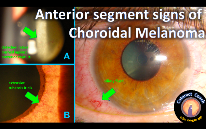
Figure 1 | At first glance, it appeared the patient had a whitish cataract (A), but, in fact, the white structure was the retina pushed up against the posterior capsule of a relatively clear crystalline lens. Further examination showed extensive rubeosis iridis (B) and hyperemia in a ciliary flush pattern (C).
A B-scan ultrasound showed a complete funnel detachment with a large mass in the vitreous cavity. MRI showed a mass that was 10 mm x 15 mm in dimension, bilobed, and attached to the scleral wall (Figure 2). After consultation with a colleague specializing in ocular oncology, I confirmed that the diagnosis was choroidal melanoma. Surgery to enucleate the eye was performed, and histopathology showed an aggressive tumor that had begun to invade the scleral wall.
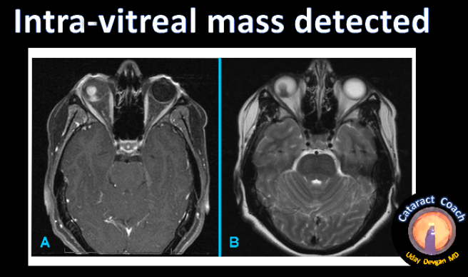
Figure 2 | MRI showed a bright lesion inside the vitreous cavity of the patient’s right eye on the TI image (A), which appears dark on the T2 image (B). The bilobed lesion was approximately 10 mm x 15 mm in size and attached to the scleral wall. Fortunately, no brain lesions were detected.
A TEAM APPROACH
In this case, the patient was completely shocked by the diagnosis, and justifiably so, as he was young and otherwise healthy. Hours were spent in detailed discussion with the patient and his family. We made all efforts to convey the seriousness of the diagnosis while remaining mindful of the patient’s psychological state. This case emphasized to me the importance of a team-based approach involving the patient’s family members and other specialists and the need to dedicate the appropriate time to fully address the situation.
Fortunately, the patient is doing well, and the systemic workup and consultation with a medical oncologist did not reveal metastasis of the melanoma. Although choroidal melanoma is rare, the loss of vision can lead to reduced mobility and independence, and it can have detrimental effects on a patient’s affect and outlook on life.
EMPATHY GOES A LONG WAY
As physicians, we feel for our patients, and we try our best to see things from their perspective. But sometimes, there is no explanation as to why a patient is struck with a grave diagnosis or a complication from surgery. One of the most important things we can do in these situations is to empathize with the patient and say, “I’m sorry you’re in this tough situation, but know that I am here to help you any way that I can.” We should remember that, one day, we will all likely be in their shoes, listening to our physicians explain the diagnoses, risks, or prognoses of our own medical and surgical challenges.
